Fig. 1
Note the equinus and varus deformity. The smaller calf of the affected side is evident
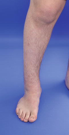
Fig. 2
Distal tibial varus deformity showing a plantigrade foot through subtalar compensation
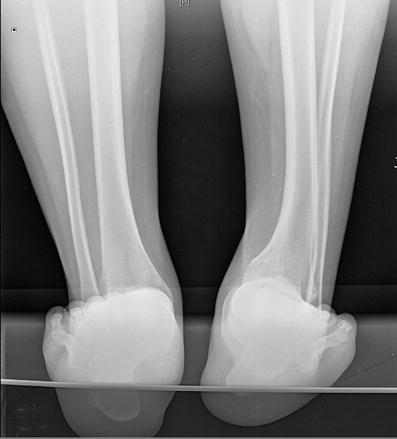
Fig. 3
Saltzman view showing the distal tibial varus
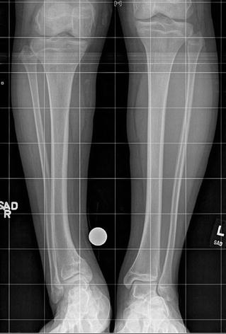
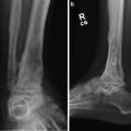
Fig. 4
AP tibias showing the right distal varus
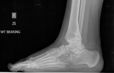
Fig. 5
Distal tibial procurvatum
3 Preoperative Problem List
Distal tibial varus-procurvatum deformity
Tibial and peroneal nerve at risk
3.5 cm tibial shortening
4 Treatment Strategy
Supramalleolar osteotomy (SMO) to correct deformity
Peroneal nerve release to prevent injury
Anterior compartment fasciotomy to prevent compartment syndrome
Tarsal tunnel release
Gastrocnemius recession (Vulpius)
Proximal tibial osteotomy: for lengthening
External fixation to allow for gradual correction of deformity and shortening
5 Basic Principles
A single-level distal tibial TSF to correct the varus-procurvatum and shortening could have been an option. However, in order to decrease the stress on the soft tissues and the ankle joint, a double-level deformity correction was performed. The lengthening through the proximal osteotomy stretches only the gastrocnemius. On the other hand, lengthening through a distal tibial osteotomy stretches all tendons around the ankle joint, the joint itself, and the neurovascular bundle. In cases of equinus and varus correction, an Achilles lengthening procedure and tarsal tunnel release have to be performed prior to the external fixator placement. For a double-level tibial osteotomy, it is recommended to perform a prophylactic anterior compartment fasciotomy.
6 Images During Treatment
See Figs. 6, 7
 8: Femoral Bone Defect
 11: Bone Transport Over a Nail for Infected Tibial Nonunion and Bone Defect
 22: Bone Transport to a Knee Fusion and Secondary Intramedullary Nailing s/p Gunshot Wound
 39: Ilizarov Ankle Fusion
 84: Lapidus Fusion with External Fixation
 65: Closed Correction of Club Foot with Ilizarov


Related posts:
8: Femoral Bone Defect
11: Bone Transport Over a Nail for Infected Tibial Nonunion and Bone Defect
22: Bone Transport to a Knee Fusion and Secondary Intramedullary Nailing s/p Gunshot Wound
39: Ilizarov Ankle Fusion
84: Lapidus Fusion with External Fixation
65: Closed Correction of Club Foot with Ilizarov
Stay updated, free articles. Join our Telegram channel
Full access? Get Clinical Tree
