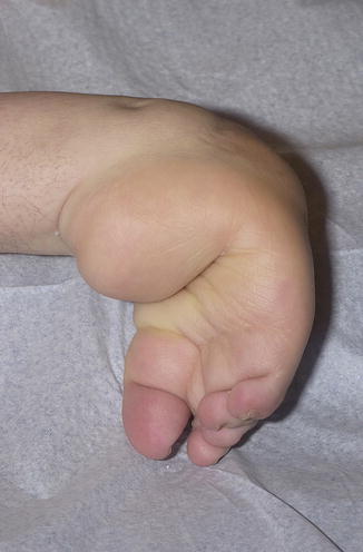
Fig. 1
Clinical view of the right foot showing a complex deformity. The left side after correction of a similar deformity in which a degree of varus and equinus deformity remained

Fig. 2
Clinical view from lateral

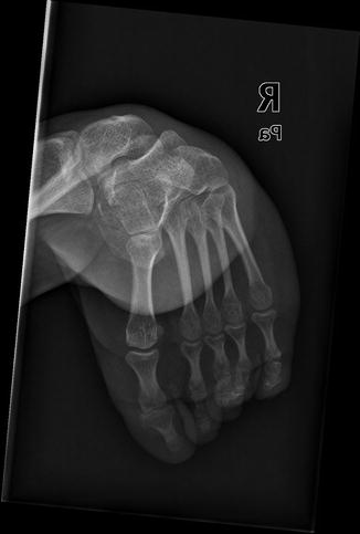
Fig. 3
X-ray lateral view of the right ankle joint showing a complex foot deformity

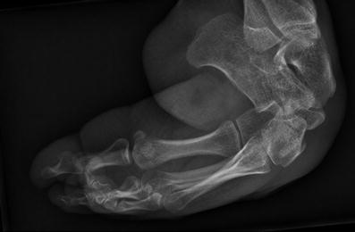
Fig. 4
Radiographical lateral view of right forefoot before treatment
3 Preoperative Problem List
On both sides a severe talipes equinocavovarus deformity existed in the foot which could not be corrected actively or passively. Especially, the soft tissues at the medial and plantar side of both feet appeared to be contracted. On X-ray bony deformities were seen at the talar, navicular, and cuneiform bones.
4 Treatment Strategy
The first step in treatment was fixing two rings to tibial bone with four K-wires under 130 kg tension followed by securing a half ring to the calcaneus by two K-wires with olives after 90 kg tensioning. From medially and laterally, two K-wires with olives were drilled through the distal and mid shaft of the metatarsal bones including a third one through their proximal ends to fixate the forefoot to another half ring after 50 kg tensioning. The setup of connections between this half ring and the calcaneal one depended on the direction and amount of the desired correction. An osteotomy was made through the talar and calcaneal bones with placing of hinges at both sites of the osteotomy. By changing orientation of hinges and using graduated rods, the forefoot half ring could be rotated gradually, correcting the supine position of the forefoot. The next step was correcting a cavus and adduction deformity by changing the orientation of hinges and graduated rods connecting the forefoot and distal tibial rings.
Seven weeks later, the forefoot alignment was restored. The next step involved reduction of the almost 90° equinus deformity of the hindfoot by gradual traction on the forefoot ring towards the most distal tibial ring by using threaded rods and hinges placed into the flexion-extension axis of the ankle joint after an open calcaneal tendon lengthening.
On this way, the equinus position could be reduced to 25°, and the slight varus position of the calcaneus was accepted. After 4 months, the external fixator was removed followed by a below-knee plaster cast for 3 weeks, permitting gradual weight bearing. Finally, orthopedic shoes were given with an adjustment to the bony swelling at the right talonavicular region which resulted into pain-free walking until now, 7 years later. Similar to the left side, a pantalar arthrodesis is expected to be the final result of treatment.
5 Basic Principles
Much attention was given to the response of the soft tissues of the foot to the distraction rate of three times a day 0.25 mm in terms of pain, skin structure and color, and signs of possibly neurological of vascular problems.
Stay updated, free articles. Join our Telegram channel

Full access? Get Clinical Tree


