Fig. 1
Lateral view
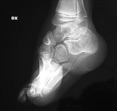
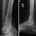
Fig. 2
Lateral radiograph before treatment
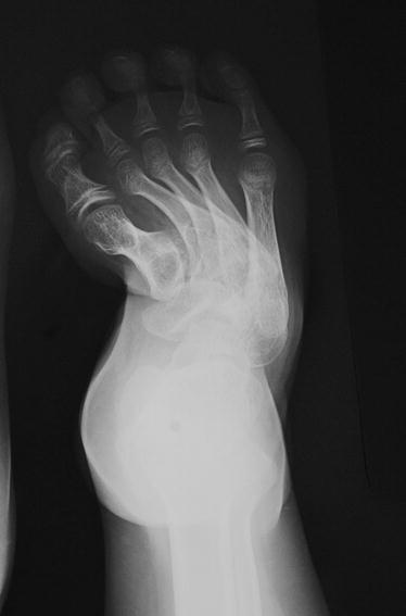
Fig. 3
Forefoot radiograph before treatment
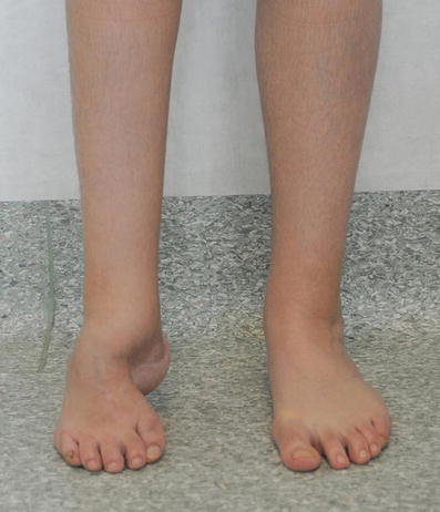
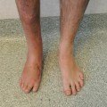
Fig. 4
Frontal view of both feet
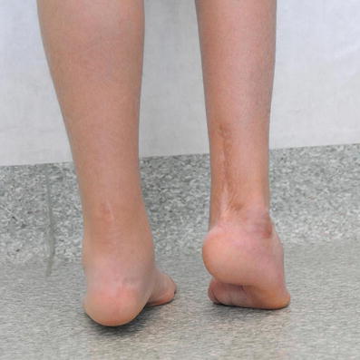
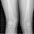
Fig. 5
Posterior view of the both feet
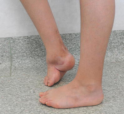
Fig. 6
Medial side view of the right foot
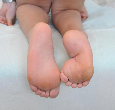
Fig. 7
Plantar view of both feet demonstrates adduction and the length discrepancy
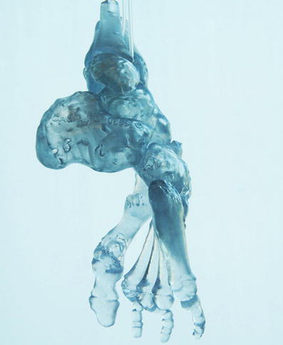
Fig. 8
Plastic model of the foot bones reconstructed on the base of 3D CT scan before treatment, lateral view
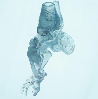
Fig. 9
Plastic model , medial view demonstrates equinus position of the first metatarsal
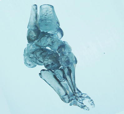
Fig. 10
Plastic model , frontal view demonstrates adduction of forefoot bones
3 Preoperative Problem List
(a)
Equinus deformity of the foot
(b)
Varus deformity of the calcaneus
(c)
Cavus deformity of the midfoot
(d)
Adduction and supination of the forefoot
(e)
Shortening of entire foot
4 Treatment Strategy
(A)
Closed correction of the deformity by distraction of soft tissue and restoration of the normal relationship between the bones of the foot.
(B)
Simultaneous correction of all components of the deformity: cavus deformity, adduction and supination of the forefoot, varus, equinus, and adduction of calcaneus.
(C)
The aim is to obtain an overcorrection of all the components of deformity and achieve a flat, dorsiflexed, abducted, and supinated forefoot.
5 Basic Principles
Gradual lengthening of the soft tissue and scars produced by external fixation utilizes the principle of tension stress which is dosed in force, power, and time (Ilizarov 1990). The goal is to create in the pathologic foot of young patients the space necessary for normal growth of the bones of the foot and the plantigrade position needed for improving joint function. The fixator achieves a multiplanar controlled correction with hinged frame and rod positioned in the line and direction of maximum stretching of soft tissue (Grill and Franke 1987
 8: Femoral Bone Defect
 11: Bone Transport Over a Nail for Infected Tibial Nonunion and Bone Defect
 22: Bone Transport to a Knee Fusion and Secondary Intramedullary Nailing s/p Gunshot Wound
 39: Ilizarov Ankle Fusion
 63: Residual Clubfoot: Equinovarus Deformity/Knee Valgus/Limb Length Discrepancy
 71: V Osteotomy of the Foot


Related posts:
8: Femoral Bone Defect
11: Bone Transport Over a Nail for Infected Tibial Nonunion and Bone Defect
22: Bone Transport to a Knee Fusion and Secondary Intramedullary Nailing s/p Gunshot Wound
39: Ilizarov Ankle Fusion
63: Residual Clubfoot: Equinovarus Deformity/Knee Valgus/Limb Length Discrepancy
71: V Osteotomy of the Foot
Stay updated, free articles. Join our Telegram channel
Full access? Get Clinical Tree
