Katarina R. Kesty and David J. Goldberg Skin Laser and Surgery Specialists of New York & New Jersey, Hackensack, NJ, USA Chronic sun exposure leads to damage of the epidermis and dermis over time, resulting in rhytides, dyschromia, alterations in skin texture, and skin laxity. In recent years there has been a progressive movement toward nonsurgical interventions for facial rejuvenation. Ablative resurfacing with CO2 and erbium‐doped yttrium aluminum garnet (Er:YAG) lasers were first used to treat photodamaged skin in the 1980s and remain the gold standard today [1–5]. Yet despite their effectiveness at treating photoaging, there has been a recent push for non‐ablative treatment modalities with less downtime and fewer side effects. There has also been an increased demand for rejuvenation in patients with darker skin types, and non‐ablative lasers have been shown to be safe for this population [6]. Non‐ablative lasers have been used to successfully treat rhytides, dyspigmentation, vascular changes, skin texture, laxity, and scarring [1, 4, 7]. The main goal of these systems is to selectively induce dermal damage, resulting in collagen remodeling and production while sparing the epidermis [1, 2,7–9]. Many different systems have been used with this endpoint in mind (Table 52.1), and they will be discussed in detail below. Non‐ablative rejuvenation systems are composed of lasers, light sources, and radiofrequency devices. Laser systems use energy in the infrared or near infrared spectrum to target specific dermal chromophores, such as water, melanin, and hemoglobin. In contrast, radiofrequency devices produce heat in the dermis and subcutaneous tissue as a result of resistance to current flow through the tissue. Although the mechanisms of action are different, the end result is the same. Both the light sources and radiofrequency devices effectively heat the dermis eliciting a wound healing response without disturbing the integrity of the epidermis [1, 4,10–12]. One of the main effects of photodamage and aging is a reduction in dermal collagen, and this is the target of non‐ablative rejuvenation. It has been proven that thermal induced injury to the dermis causes local release of inflammatory cytokines leading to the proliferation of fibroblasts which results in collagen synthesis [813–16]. In vivo mouse studies have been performed to evaluate collagen composition after treatment with various lasers, and they have all demonstrated a significant change in collagen and extracellular matrix [8, 14]. Improvement of the cosmetic appearance of the face depends on the ability to restore damage done over time due to sun exposure. This includes improving red and brown areas, improving skin texture and tone, and reducing the number and depth of rhytides. Many non‐ablative systems have been evaluated for efficacy in facial rejuvenation. Results have been promising and often require multiple treatments in order to appreciate clinical improvement. There are many non‐ablative laser regimens that are effective for facial rejuvenation, and the corresponding studies for each laser will be discussed below. The most common lasers and parameter settings used today are outlined in Table 52.2. Table 52.1 Lasers for facial rejuvenation. The 532 nm KTP laser is one of the many modalities used in facial rejuvenation. It is absorbed more intensely by melanin and hemoglobin, and so it is thought that fewer treatments are needed when targeting these components of photodamaged skin [3]. This laser has been shown to effectively target vascular and pigmentary change, skin texture, tightening, acne scars, and rhytides [4, 16]. Lee performed a 150‐person study to compare the efficacy of the KTP laser alone, the long pulsed Nd:YAG alone, or the two modalities combined on collagen enhancement and photorejuvenation [16]. After three to six treatments, all three groups showed statistically significant improvement in rhytides, skin tone and texture, reduction in redness, and improvement in dyschromia. Overall, the KTP laser treated patients showed more improvement than the LP Nd:YAG laser, but the combined treatment group was superior to either group alone. The combined group had a 70–80% improvement in redness and pigmentation, 40–60% improvement in skin texture, tone and tightening, and a 30–40% improvement in rhytides. In addition to these findings, it was also noted that collagen remodeling continues for up to 6–12 months posttreatment, with slow regression in benefit thereafter. Although further studies are necessary, these results suggest that combined modalities may be superior at targeting various factors involved in photodamage and more effectively stimulate collagen production. A study by Ho and colleagues compared the effectiveness and safety of using the 532 nm LP KTP laser, 595 nm long pulse dye laser (LPDL), 755 nm LP Alexandrite laser, and 532 nm QS Nd:YAG laser for the treatment of freckles or lentigines in Fitzpatrick skin type III and IV Asian patients [17]. Forty patients were enrolled in the study and each patient attended between one and four treatments at 4–6 weeks intervals depending on the clinical response. The laser parameters included a fluence of 12–14 J/cm2 and spot size of 2 mm for the LP KTP laser, a fluence of 11–13 J/cm2 and spot size of 7 mm for the LPDL, a fluence of 20–35 J/cm2 and spot size of 10 mm for the LP Alexandrite laser, and a fluence of 0.6 J/cm2 and spot size of 2 mm for the QS Nd:YAG laser. Statistically significant improvement of global and focal facial pigmentation was found after treatment with LP KTP, LPDL, and QS Nd:YAG lasers. No significant improvement was found after treatment with LP Alexandrite laser. Post‐inflammatory hyperpigmentation was absent after LP KTP and LPDL treatment, but was seen in 20% of patients after LP Alexandrite treatment and 10% of patients after QS Nd:YAG treatment. The 532 nm LP KTP and 595 nm LPDL lasers appear to be more effective with less complications compared to the 532 nm QS Nd:YAG and 755 nm LP Alexandrite lasers for the treatment of freckles and lentigines in Fitzpatrick type III and IV skin. The authors also concluded that a long pulse laser and small spot size appear to reduce the risks of lentigines treatment in darker skin types. Another recent study by Negishi et al. evaluated 532 nm KTP laser alone or in combination with LP 1064 to treat dyschromia in 22 Japanese patients. These patients were treated with three sessions 2 weeks apart with one side of the face with 532 and the other with 532 and LP 1064. 532 was shown to be a safe and effective treatment and the addition of 1064 was not clinically apparent but a difference was detected by the 3D skin analysis device [18]. Table 52.2 Non‐ablative lasers. The pulse dye laser (PDL) is a yellow light that targets oxyhemoglobin and melanin. It has been used to treat vascular changes and induce new collagen formation. It has been hypothesized that wavelengths targeting hemoglobin disrupt vascular endothelial cells resulting in cytokine release and subsequent collagen remodeling and production [4, 19, 20]. A group of 10 female patients, Fitzpatrick types I–IV, were treated in the periorbital area with the 595 nm flash lamp PDL [20]. One side of the face was treated with the following laser settings: 1.5 ms pulse, fluences of 5–6 J/cm2, and a spot size of 7 mm. The contralateral side was treated with a 40 ms pulse duration, fluences of 8–11 J/cm2, and a 7 mm spot size. Cryogen cooling was administered for 30 ms with a delay of 30 ms before treatment. Subjects were treated one to two times, and at their 6‐month follow‐up, 70% treated had mild to moderate improvement overall. Sixty percent had equal improvement on both sides despite the different settings. Histologic and electron microscopic evaluation showed a significant increase in papillary dermal collagen, mainly type I. Another study performed by Bernstein evaluated the effects of treatment of sun damaged skin with a 595 nm long pulse duration PDL [19]. Ten subjects were treated with 10 ms pulse duration, fluences of 8–10 J/cm2, and a 10 mm spot size. Improvements were evaluated 8 weeks after treatment with photograph comparison. Blinded physician evaluation of pre‐ and post‐photographs rated improvement in wrinkles in 50%, facial veins improved in 82%, overall redness improved 80%, pigmentary change 61.4%, and a 25% improvement in pore size. A small clinical trial was performed on 10 patients comparing the efficacy of a long‐pulse PDL (LPDL) versus an intense pulsed light (IPL) on photodamaged facial skin [21]. When compared to the IPL, the LPDL showed greater improvement in lentigines (81% versus 62%), no significant difference in wrinkle reduction between the two groups, and fewer treatments were needed with the LPDL when compared to the IPL (3 versus 6). In addition to using the PDL laser as a single agent in photorejuvenation, recent trends have shown an effective combination with aminolevulinic acid (ALA) in treatment of photodamaged skin. It is believed that PDL at 595 nm activates protoporphyrin IX, a photosensitizer, which accumulates in photodamaged cells, causing destruction of the cells, release of cytokines, and collagen repair [1]. This is a new and exciting area of research that offers another effective treatment modality for photorejuvenation. The IPL system emits light, with wavelengths of 550–1200 nm, which effectively targets melanin, hemoglobin, and water (to a lesser degree). The IPL device has been used in photorejuvenation to target vascular changes, pigmentary alteration, and mild rhytides (Figures 52.1–52.3) [1, 3, 4, 21, 22]. Filters may be used with the IPL to target specific chromophores. Rapid improvement in overall appearance after IPL treatment is secondary to rapid and effective improvement of vascular and pigmentary change, rather than improvement in wrinkles [4]. As per Weiss et al., shorter pulse duration and lower cut off filters when using the IPL system, results in significant improvement in pigmentary alteration [3]. Although not thought to be the most effective treatment modality for rhytides, some studies using the IPL have shown improvement [1, 2, 5, 21, 22]. Goldberg et al. evaluated the treatment of facial rhytides with an IPL system using a 645 nm cutoff filter [2, 22]. Thirty patients, skin type I–III, were treated one to four times over a 10‐week period. Treatments were delivered using fluences of 40–50 J/cm2, through bracketed cooling device with triple 7 ms pulses, and interpulse delay of 50 ms. At 6 months follow‐up, approximately 53% showed some improvement, 30% showed substantial improvement, and 17% showed no improvement. Figure 52.1 Forty‐four‐year‐old female before and after five treatments with intense pulse light for facial rejuvenation. Figure 52.2 Forty‐five‐year‐old before and after two treatments of intense pulse light to the chest for dyschromia. Figure 52.3 Forty‐five‐year‐old female before and after two treatments with intense pulse light to the face for rejuvenation. Hedelund et al. also performed a study looking at the efficacy of IPL treatment for perioral rhytides in comparison to CO2 ablative resurfacing [5]. Twenty‐seven females, skin type II, with perioral rhytides were randomly treated with 3 monthly IPL sessions or one CO2 laser ablation. The results showed a higher degree of patient satisfaction and significant improvement in rhytides with CO2 laser resurfacing when compared to IPL rejuvenation. In addition to the more dramatic improvement seen with CO2 resurfacing, side effects were also found to be significantly higher in this group. Patients treated with the CO2 laser experienced milia, dyspigmentation, and persistent erythema, while the IPL group wasn’t noted to have any side effects. Both groups showed long term improvement in skin elasticity, although no significant improvement in wrinkles was seen in the IPL treatment group. Many physicians today advocate three to six treatments for significant improvement, which implies that the treatment course may not have been sufficient to produce notable results. Another study by Barikbin et al treated 38 females with 3 sessions of monthly IPL for periocular rejuvenation [23]. After 6 months, approximately 85% of patients noticed improvement in the appearance of their periocular area. One study treated 10 Asian females with 5 monthly IPL treatments and found that after the treatment, dermal papillary density and thickness of the basal layer were significantly increased and mean capillary diameter was significantly reduced [24]. As with PDL lasers, recent trends have shown effective combination of IPL with ALA in treatment of photodamaged skin [25–28]. Patients treated with this combinational therapy demonstrated greater improvement in global photodamage, mottled pigmentation, and fine lines compared to those patients treated with IPL alone [25]. Combinational treatment is well tolerated with little difference in the incidence of adverse effects with or without 5‐ALA pretreatment. IPL can be combined with other treatments for optional facial rejuvenation. One study examined the combination of IPL, bipolar radiofrequency, and infrared diode laser administered at the same visit for four visits 1 month apart [29]. This combination improved erythema, telangiectasias, and skin texture for up to 6 months after treatment with a high degree of patient satisfaction. In another study of 71 patients, IPL was combined with non‐ablative 1410 and non‐ablative RF to improve periocular aging [30]. This combination was well tolerated and showed the greatest improvement in skin elasticity and to a lesser extent skin erythema and pigmentation. One of the first laser systems to be developed for non‐ablative rejuvenation was the Nd:YAG 1320 nm laser [31]. At this wavelength, energy is able to penetrate into the papillary and mid reticular dermis, and it is absorbed by water associated with dermal collagen. There is a high water absorption and strong scattering in the dermis which allows for extensive dermal wounding [31]. This accelerates the productive capacity of fibroblasts as seen in its promotion of the two major secretory factors they produce: basic fibroblast growth factor (bFGF) and inhibiting transforming growth factor β1 (TGF‐β1) [32, 33]. This is followed by the stimulation of collagen types I, III, and VII, and tropoelastin production [33, 34]. The surface cooling systems are present to protect the epidermis from involvement. The 1320 nm Nd:YAG laser has been used to target acne scarring, photoaging, and rhytides with variable results [1, 2, 31, 35]. As a result of its poor absorption by melanin, it can be used in all skin types without fear of pigmentary change [35]. When treating mild rhytides or acne scars, Weiss et al. recommends using a fluence of 17–19 J/cm2, a total of 25 ms of cooling (pre‐ and post‐cooling at 10 ms and mid‐cooling at 5 ms), with a fixed pulse duration of 50 ms, and two to three passes [3]. When treating acne scars with this regimen, 30–50% improvement has been observed in about four of five patients, while 20% show no significant response. According to a study done by Rogachefsky et al., the 1320 nm Nd:YAG laser is an effective modality to treat atrophic and mixed pattern facial acne scars [35]. After treating 12 patients with three monthly sessions, optimal improvement was noted in atrophic acne scars; however, mixed acne scars also showed softening of sclerotic and shallow pitted scars. Sadick et al. also confirms significant improvement in acne scars after treatment with the 1320 nm Nd:YAG, but he proposes that six sessions is more effective than three laser sessions [31]. The QS Nd:YAG 1064 nm laser has not only proven to be successful for treatment of tattoos, vascular, and pigmented lesions, but it has recently been used to treat rhytids, photodamage, and acne scars [2, 9, 12, 36]. This laser system is poorly absorbed by water, making deeper collagen damage more likely when compared to systems with other wavelengths [36]
CHAPTER 52
Non‐ablative Lasers
Introduction
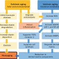
Pathophysiology
Non‐ablative modalities
Laser type
System name
KTP
ExcelV (Cutera)
NewSurg KTP (NewSurg)
Pulsed dye 585/595 nm
ADVATx (Advalight) (589 nm)
VBeam Perfecta (Candela)
Spectra (Lutronic)
DenaVe (Quanta/Cartessa)
Intense pulse light (IPL)
Nordlys (Candela)
JouleX (Sciton)
Stellar M22 (Lumenis)
Icon (Cynosure)
Xeo (Cutera)
Venus Versa (Venus Concepts)
Optimas (InMode)
Luxea (DEKA/Cartessa)
1320 nm Nd:YAG
ADVATx (Advalight) (1319 nm)
ThermaScan (Sciton)
1064 nm QS‐Nd:YAG
Stellar M22 (Lumemis)
MedLite C6 (Cynosure)
Spectra (Lutronic)
ClearLift (Alma)
532, 755, and 1064 nm picosecond
Picosure (Cynosure)
PicoWay (Candela)
PiQo4 (Lumenis)
PicoPlus (Lutronic)
Enlighten (Cutera)
Er:glass 1540 nm
Aramis‐Quantel (Quantel Medical)
Icon (Cynosure)
ClearSkin Pro (Alma)
1550 nm
Fraxel Restore Dual (Fraxel)
Nordlys (Candela)
Ydun (Candela)
Infrared light (1100–1800 nm)
Titan (Cutera Inc.)
1927 nm Thulium
Fraxel Restore Dual (Fraxel)
LaseMD Ultra (Lutronic)
Clear and Brilliant (Solta)
mJoule (Sciton)
Radiofrequency devices
Elos Plus (Candela)
Monopolar
Unipolar and bipolar
eTwo (Candela)
LegendPlus (Lumenis)
Venus Versa/Viva (Venus Concepts)
Optimas (InMode)
Thermage (Solta)
eMatrix (Syneron)
Potassium titanyl phosphate (KTP) 532 nm laser
Laser type
Wavelength (nm)
Fluences (J/cm2)
Pulse duration
Spot sizes (mm)
Target
KTP (pulsed)
532
7–15
20–50 ms
2, 4, 10
Hemoglobin, melanin
Pulsed dye laser (PDL)
585
3–6.5
350, 450 ms
5, 7, 10
Hemoglobin, melanin
595
6–12
6–10 ms
7, 10
Hemoglobin, melanin
IPL
550–1200
25–28
2.4, 4.0 ms
Hemoglobin, melanin, water (weak)
Nd:YAG
1320
17–22
200 or 350 ms
6, 10
Water
Nd:YAG (QS)
1064
2.5–7
5 ns
6
Hemoglobin, melanin, water
Er:glass
1540
Up to 126
3.3
4, 5
Water
1550 Er:glass nm
1550
70 mJ/MTZ
Water
Infrared light
1100–1800
30–40
170–200 pulses
–
1927 nm
1927
20 mJ/MTZ
Variable
Water
Radiofrequency
RF bipolar current
61.5–63.5 (adjusted for pain)
300–400 pulses
0.25, 1.0, 1.5, 3 cm
–
RF, monopolar
RF, unipolar
RF electromagnetic radiation
50–250
–
RF, bipolar
RF bipolar current
40–100
Pulse dye laser (PDL) 585 or 595 nm
Intense pulsed light (IPL)
1320 nm Neodymium yttrium aluminum garnet (Nd:YAG)
Q‐switched (QS) Nd:YAG 1064 nm laser
Related posts:
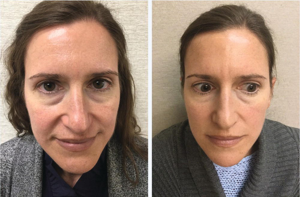
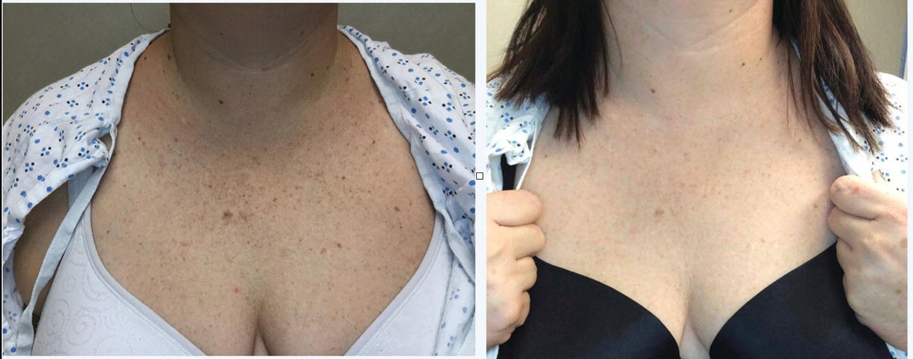
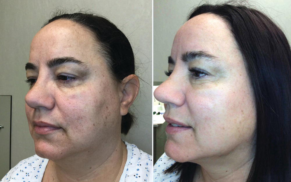
![]()
Stay updated, free articles. Join our Telegram channel


Full access? Get Clinical Tree


