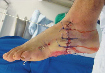
Fig. 1
The plantar aspect of the foot is seen with soft tissues and bone loss as well as massive wound contamination

Fig. 2
The talus is visible in the wound. The surrounding muscle and soft tissue are vascularized. There is bone and soft tissue instability
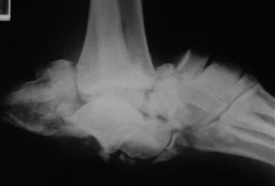
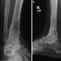
Fig. 3
The entire foot has been split in half and dislocated around the talus
3 Preoperative Problem List
Severe trauma to soft tissue and bone loss
Massive contamination and delayed transfer to the hospital
Short bone fragments and comminution
4 Treatment Strategy
Damage control and prevention of infection through thorough debridement of the bone and soft tissue, provisional bony reduction, and stabilization with K-wires and external fixation (Fig. 4). A simple external fixator is used at first to allow for repeat open debridements. The wound is closed primarily after all the damaged tissue has been removed from the zone of injury (Fig. 5).
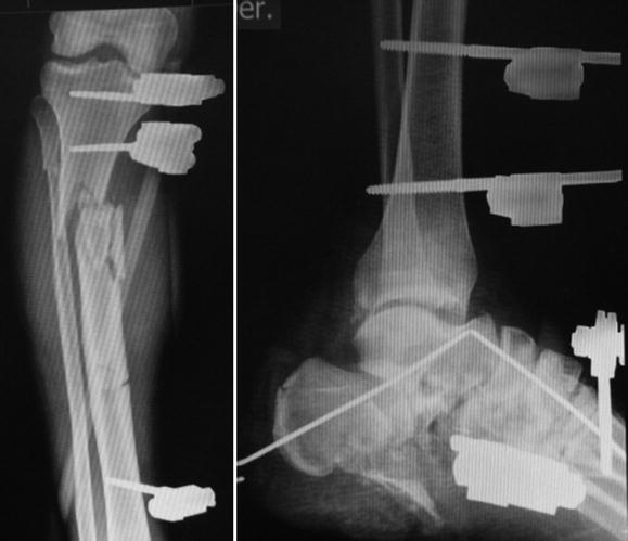
A combination treatment with the integration of internal and external fixation techniques is applied. The circular fixator is utilized to provide superior stability and comfortable patient mobilization (Fig. 6). The frame is extended proximally to control the diaphyseal tibial fracture. Percutaneous hindfoot fusion with internal fixation is implemented for definitive fixation (Fig. 7). These cannulated screws also prevent subtalar separation during correction of the equinus ankle position (Fig. 8). The tibia fracture was further stabilized with an IM nail. The use of the fixator was limited to the control of the foot fractures and the ankle joint position.
5 Basic Principles
Stabilization of bone and soft tissues.
Immediate antibiotic IV.
Rational use of Ilizarov method.
Integrated fixation: the surgeon must be careful to avoid contact between internal and external fixation to prevent infection.
6 Images During Treatment
See Figs. 4, 5, 6, and 7.
 8: Femoral Bone Defect
 11: Bone Transport Over a Nail for Infected Tibial Nonunion and Bone Defect
 22: Bone Transport to a Knee Fusion and Secondary Intramedullary Nailing s/p Gunshot Wound
 39: Ilizarov Ankle Fusion
 84: Lapidus Fusion with External Fixation
 65: Closed Correction of Club Foot with Ilizarov


Fig. 4
Soft tissue stability – monofilament sutures without tension
Fig. 5
External fixator in damage control (first step)
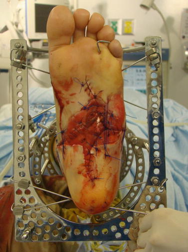
Fig. 6
Ilizarov method. Minimally invasive external fixation and dynamic stability
Related posts:
8: Femoral Bone Defect
11: Bone Transport Over a Nail for Infected Tibial Nonunion and Bone Defect
22: Bone Transport to a Knee Fusion and Secondary Intramedullary Nailing s/p Gunshot Wound
39: Ilizarov Ankle Fusion
84: Lapidus Fusion with External Fixation
65: Closed Correction of Club Foot with Ilizarov
Stay updated, free articles. Join our Telegram channel
Full access? Get Clinical Tree
