Fig. 1
Sensate and well-perfused foot
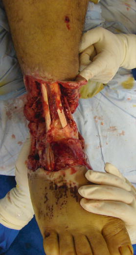
Fig. 2
Palpable dorsalis pedis pulse
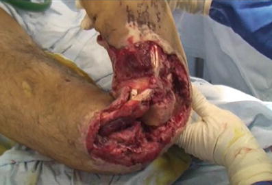
Fig. 3
Bone and soft tissues instability
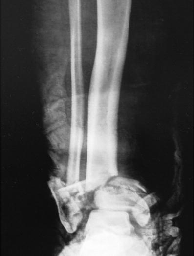
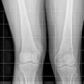
Fig. 4
AP radiograph showing bone loss and comminution
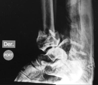
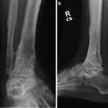
Fig. 5
Lateral X-ray showing bone and joint damage
3 Preoperative Problem List
Severe trauma to soft tissue
Massive contamination and delayed transfer to the hospital
Ankle joint destruction
Bone loss and limb shortening
4 Treatment Strategy
Transfer to a third-level multidisciplinary center.
Immediate damage control with limb stabilization and soft tissue management.
Acute shortening techniques.
Ankle fusion.
Combination of proximal tibial lengthening and internal and external fixation techniques .
5 Basic Principles
Stabilization of bone and soft tissues
Immediate antibiotic IV
Rational use of the Ilizarov method
6 Images During Treatment
See Figs. 6, 7, 8, 9, 10, 11, and 12.
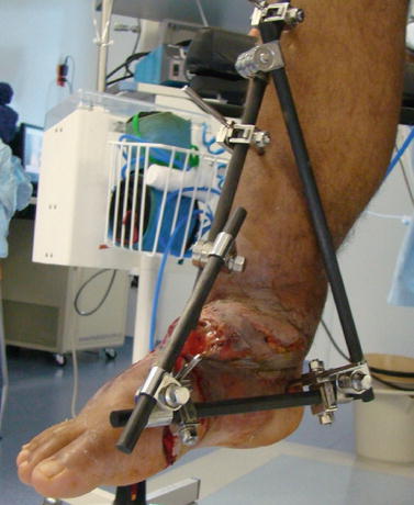
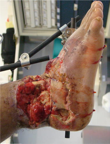
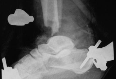
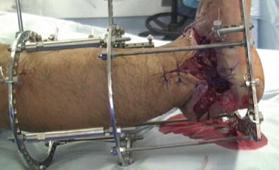
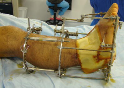
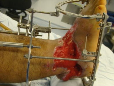
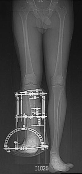
Fig. 6
Soft tissues stability
Fig. 7
After shortening and provisional stabilization with pin-to-bar fixation
Fig. 8
Lateral radiograph demonstrates bone loss after thorough debridement including resection of the distal tibia
Fig. 9
Ilizarov method. A minimally invasive technique was used to convert to a circular external fixator
Fig. 10
The skin loss was treated with a hydrophilic matrix cover
Fig. 11
Granulation tissue thrives with limb stability, shortening of the soft tissue defect, and use of the matrix cover
Fig. 12
A long, standing X-ray demonstrates severe limb shortening and bone stabilization with the Ilizarov device
Related posts:
 8: Femoral Bone Defect
 11: Bone Transport Over a Nail for Infected Tibial Nonunion and Bone Defect
 22: Bone Transport to a Knee Fusion and Secondary Intramedullary Nailing s/p Gunshot Wound
 39: Ilizarov Ankle Fusion
 63: Residual Clubfoot: Equinovarus Deformity/Knee Valgus/Limb Length Discrepancy
 65: Closed Correction of Club Foot with Ilizarov
Stay updated, free articles. Join our Telegram channel

Full access? Get Clinical Tree


