The authors present a 3D in vivo imaging system used to assess the effectiveness of IPL and fractional laser treatments of photodamaged skin. Preoperative and postoperative images of patients treated with these procedures are analyzed and demonstrate the superior ability of this 3D technology to reveal decrease in vascularity and improvement in melanin distribution and calculate the degree of individual deep wrinkles before and after treatment.
Topography of the skin surface as well as melanin and hemoglobin concentration and distribution are a mirror of the functional skin status. Changes in these features not only are a tool for early-stage diagnosis of diseases but also give an indication of the response to medical and cosmetic treatment. Therefore, their evaluation is of great interest for dermatologic research. However, although physicians can apply classification rules to visual diagnosis, the overall clinical approach is subjective and qualitative, with a critical dependence on training and experience. Over the past 20 years, several noninvasive techniques for measuring the skin’s properties have been developed and tested to extend the accuracy of visual assessment alone. Many of them were not only very precise but also very complicated; others were very simple but approximations.
In this article, a new optical, precise, user-friendly measuring system is presented to demonstrate the effectiveness of the most common laser treatments on photodamaged skin. Many of the skin changes commonly associated with aging, changes in pigmentation, telangiectasia, sallowness, and wrinkling are actually the result of sun exposure. Changes in pigmentation (blotchy brown freckles and age spots), dryness, areas of redness, thinning of the dermis, loss of elasticity, fine lines, and deep wrinkles are all signs of chronic UV exposure. Excluding injectables, surgery, and peelings, the most common procedures to improve these changes in the skin appearance are the IPL treatments and, more recently, the fractional resurfacings. Preoperative and postoperative images of patients treated with these procedures and analyzed using this new three-dimensional (D) in vivo optical skin imaging system are shown.
The skin imaging device
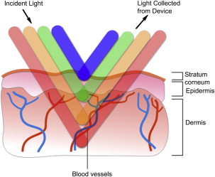
The Antera 3D (Miravex, Ireland) imaging system consists of a handheld imaging device connected through a long firewire cable to a computer. The system is completed by proprietary software running on a Windows-based standard laptop or desktop personal computer. The imaging technique of Antera 3D is based on the acquisition of a number of images under varying but strictly controlled illumination conditions. Several light-emitting diodes are used to illuminate the skin with different colors and different illumination directions ( Fig. 1 ). The acquired image data are then used for spatial and spectral analysis for reconstruction of the texture of the skin and analysis of skin constituents.
Skin texture reconstruction is achieved using a technique based on shape from shading, substantially modified to eliminate skin glare and vastly improve the accuracy of measured data. The texture reconstructed in this way is then used for quantitative skin analysis, such as depth and width of wrinkles, lesions of the skin, and overall skin roughness.
The acquired spectral data are used to map the distribution and concentration of melanin and hemoglobin. Unlike traditional imaging techniques, in which only 3 color channels (red, green, and blue) are used, the Antera 3D uses reflectance mapping of 7 different light wavelengths spanning the entire visible spectrum. This mapping allows for a much more precise analysis of the skin colorimetric properties, which are mostly determined by 2 dominant chromophores: melanin and hemoglobin. Acquired spectral images are transformed into skin spectral reflectance maps, and the skin surface shape is used to compensate for light intensity variation due to the varying direction of incident illumination. The reflectance data are transformed into skin absorption coefficients and used to quantify melanin and hemoglobin concentrations using mathematical correlation with known spectral absorption data of these chromophores.
The images acquired with the Antera 3D can be visualized in several different modes: standard color skin, texture elevation map, and melanin and hemoglobin concentration maps with 2D and 3D perspective representation. The clinician can select specific skin areas for quantitative analysis and carry out before and after analyses with previously acquired images. Spot-On, the automatic matching technique that registers two or more images to one another is used to compensate for relative shifts and rotations between images, ensuring accurate data analysis. The measurement data can be presented by quickly creating a report that shows the analyzed images together with the measured values and comparison charts. Data can be stored on the computer or included in other document processing applications such as Microsoft Word or Microsoft PowerPoint.
Numeric data collected from one image must not be considered as absolute values but 2 or more images must be compared. The percentage modification of these data (data on melanin distribution, hemoglobin distribution, and surface topography) is very useful to demonstrate the effectiveness of a treatment.
Intense pulsed light treatment of facial aging skin
Current trends in aesthetic treatment of facial skin call for an effective adjunct to injectables or surgery. Patients look for treatment that offers a return to a more youthful appearance through restoration of even color and smoothness, relief from pigmentary sun damage, and the redness associated with ectatic vessels. In addition, this patient group requires treatments that are short and pain free and allow immediate return to all social activities.
Following more than 20 years of treatment of vascular lesions using the pulsed dye laser, new laserlike intense pulsed light (IPL) devices were developed at the end of the last century. These IPL devices treat these UV exposure–correlated conditions with success and provide a solution for the essential lifestyle criteria when used in a carefully administered program. This new IPL skin rejuvenation technique now has a clinical history of more than 300,000 treatments with excellent patient acceptance. IPL differs from laser light in that, rather than monochromatic single wavelength, IPL emits a noncoherent broad-spectrum light. The IPL devices used in the rejuvenation procedure emit a spectrum extending from 500 nm to 1200 nm. To customize the light energy delivery for a given procedure, the operator uses a cutoff filter, or light guide, of designated wavelength, below which the spectrum is selectively eliminated. The IPL system conforms to the principle of selective photothermolysis. For dilated vessels, as seen in patients with sun damage and rosacea, the light energy with high absorption by hemoglobin and oxyhemoglobin reaches the dermal capillary bed and selectively destroys the abnormal vessels. For sun spots and lentigos, as seen in patients with sun damage, the light crumbles the granules of melanin distributed at the dermoepidermal junction. Macrophages can then therefore remove these smaller granules of melanin. The operator controls all aspects of the light pulse, including cutoff wavelength (nm), energy level (J/cm 2 ), pulse duration (milliseconds), pulse pattern (single, double, or triple), and delay time between pulses (milliseconds). This allows for precise control of light energy, which in this procedure is used for customization for skin type, procedure progress, and other variables.
Big vessels of the nose can be treated ( Fig. 2 ) with few sessions of treatment combining different cutoff (590and 560 nm), different energy (22 and 19 J/cm2), and different pulse duration and delay time. The 3D images ( Fig. 3 ) visually clarify the effectiveness of the treatment while the report that mathematically analyses the prepictures and postpictures presents the percentage of improvement ( Fig. 4 ).