Disorders of the Genitalia, Perineum, and Anus 
 Anogenital skin and mucosa are subject to unique disorders because of their special anatomy.
 Dermatologic and systemic disorders occur in the anogenital region.
 Primary neoplasms arise in these areas, most commonly associated with chronic human papillomavirus (HPV) infection.
 Sexually transmitted as well as other infections also occur commonly in these sites.
 Often normal structures, newly observed, give rise to great concerns about sexual transmitted infections such as anogenital warts and molluscum contagiosum.
Pearly Penile Papules ICD-9: 607.89 ° ICD-10: N48.89 
 Normal anatomic structures. Incidence: Up to 19%.
 Symptoms: Asymptomatic; may arouse some anxiety when first noted.
 Clinical findings: Skin-colored 1- to 2-mm, discrete, domed papules evenly distributed circumferentially around the corona (Fig. 34-1), giving a cobblestone pattern.
 Differential diagnosis: Condylomata acuminatum, molluscum contagiosum.
 Histology: Angiofibromas.
 Management: Reassurance: normal anatomic structures.
 Synonym: Angiofibromas.
Figure 34-1. Pearly penile papules Pink (skin-colored), 1- to 2-mm papules are seen regularly spaced along the corona of the glans penis. These structures, which are part of the normal anatomy of the glans, are commonly mistaken for condylomata or molluscum contagiosum.
Sebaceous Gland Prominence ICD-9: 789,9 ° ICD-10: Q89.9 
 Normal sebaceous glands. Analogous to sebaceous gland on mucosa of mouth.
 Locations: Penis, vulva.
 Manifestation: 2-mm dermal papule; cream colored. May be arranged in rows.
 Synonyms: Tyson glands, sebaceous hyperplasia, “ectopic” sebaceous glands, Fordyce condition.
Angiokeratoma (See also Section 9) 
 Ectatic thin-walled blood vessels in the superficial dermis with overlying epidermal hyperplasia.
 Increasingly common with aging.
 Multiple purple, smooth, 2- to 5-mm papules. Bleed with trauma. (See Section 9, Fig. 9-25).
 Location: Scrotum, glans penis, penile shaft. Labia, vulva.
 Differentiate from angiokeratomas of Fabry disease (usually pinhead size, found on bathing trunk area and upper thighs), Kaposi sarcoma.
 Management: Reassurance, electrosurgery.
 Synonym: Angiokeratomas of Fordyce.
Sclerosing Lymphangitis of Penis ICD-9: 607.2 ° ICD-10: N48.29 
 Etiology Trauma associated with vigorous sexual activity.
 Pathogenesis: Lymphatic stasis may result in thrombosed lymphatic vessels. Subsequent recanalization and fibrosis of walls of lymphatic vessels.
 Clinical findings: Painless, firm, at times nodular, translucent serpiginous cord appears suddenly,
 usually parallel to corona; not attached to overlying epidermis (Fig. 34-2).
 Course: Resolves spontaneously in weeks to months.
 Synonyms: Nonvenereal sclerosing lymphangitis, penile venereal edema, Mondor phlebitis.
Figure 34-2. Sclerosing lymphangitis: penis A dermal cord on the distal shaft parallel to the corona.
Lymphedema of the Genitalia ICD-9: 457.1 ° ICD-10: 189.0 
 Acute idiopathic scrotal edema. Occurs in young boys. Resolves spontaneously in 1-4 days. Differentiate from acute scrotum. Also reported in adults with dengue hemorrhagic fever, Henoch-Schönlein purpura.
 Lymphogranuloma venereum (see Section 30). Occurs in chronic undiagnosed infection. Both sexes. Referred to as esthiomene: elephantiasis due to lymphatic obstruction. Chronic. Deformity of penis referred to as “saxophone penis.”
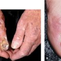
 Chronic recurrent bacterial infection may be causative (Fig. 34-3A, B).
 Kaposi sarcoma.
 Filarial or lymphatic elephantiasis. Caused by parasitic worms such as Wuchereria bancroftii, Brugia malayi, B. timori. Associated with elephantiasis of legs.
 Synonym: Lymphangiofibrosis thrombotica occlusiva.
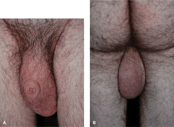
Figure 34-3. (A, B) Chronic lymphedema: scrotum A 29-year-old male with history of recurrent scrotal infections that have destroyed lymphatic channels. There is scrotal noncompressible lymphedema and the penis is retracted.
Plasma Cell Balanitis and Vulvitis 
 Asymptomatic red glistening plaque(s) on glans penis (Fig. 34-4) or vulva.
 Differentiate from squamous cell carcinoma in situ.
 Management: Circumcision is curative in uncircumcised males. Otherwise, topical corticosteroids, calcineurin inhibitors, and imiquimod can be used. Electrosurgery and laser destruction have also been reported.
 Synonym: Zoon balanitis.
Figure 34-4. Plasma cell balanitis Solitary red glistening plaque for 10 years in an uncircumcised male.
Phimosis, Paraphimosis, Balanitis Xerotica Obliterans ICD-9: 607.81 ° ICD-10: N48.0 
 Phimosis: nonretractable foreskin. Etiology: Lichen sclerosus, nonspecific balanoposthitis (posthitis is inflammation of foreskin or prepuce), lichen planus, cicatricial pemphigoid, chronic lymphedema, Kaposi sarcoma. Precludes examination of glans for precancerous changes (Fig. 34-5).
 Balanitis xerotica obliterans (BXO): End stage of chronic phimosis. Foreskin fibrotic, contracted, fixed over glans and cannot be retracted over glans. Most often end-stage lichen sclerosus, which is commonly referred to as BXO (see Section 14, lichen sclorosus).
 Paraphimosis: Foreskin fixed in retraction. Etiology: vigorous sexual activity, acute contact urticaria, acute allergic contact dermatitis, lichen sclerosus (Fig. 34-6).
Figure 34-5. Phimosis The prepuce or foreskin has been chronically inflamed with scarring and is no longer retractable over the glans penis.
Figure 34-6. Paraphimosis The prepuce or foreskin has been retracted proximally over the glans and cannot be replaced to the normal position covering the glans. The shaft is edematous.
Genital (Penile/Vulvar/Anal) Lentiginoses ICD-9: 709.8 ° ICD-10: L98.8 
 Onset: Adulthood.
 Clinical findings: Tan, brown, intense blue-black; usually variegated, 5- to 15-mm macules.
 Sites: In clusters on vulva (labia minora, Fig. 34-7), penis (glans, shaft) (Fig. 34-8), and perianal areas.
 Course: Persist for years without change in size.
 Histology: No significant melanocytic hyperplasia; nevus cells are not present; pigmentation due to increased melanin in basal cell layer.
 Differential diagnosis: Melanoma in situ, PUVA lentigo, fixed drug reaction, blue nevus, HPV-induced intraepithelial neoplasia (IN).
 Diagnosis: Dermoscopy rules out in situ melanoma; histology confirms diagnosis.
 Extensive lesions that cannot be easily removed should be followed photographically; areas that show significant change should be biopsied.
 Synonyms: Penile lentigo, vulvar melanosis.
Figure 34-7. Genital lentiginoses: vulva Multiple, variegated dark brown macules, bilaterally on the labia minora. Acrolentiginous melanoma in situ must be ruled out.
Figure 34-8. Genital lentiginoses: penis Variegated macular pigmentation of the glans and foreskin for over 20 years. Biopsy ruled out melanoma and HVP-infection (SCCIS).
Vitiligo and Leukoderma (See also Section 13) 
 Etiology: Loss of melanocytes results in depigmentation.
 Isomorphic or Koebner phenomenon: Depigmentation at sites of injury: genital herpes, cryosurgery, imiquimod therapy.
 Wood lamp examination: Differentiates depigmentation from hypopigmentation.
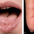
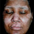
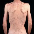
 Clinical findings: Sharply demarcated, depigmented, white macules (Fig. 34-9); examine skin for other depigmented areas.
 Differential diagnosis: Lichen sclerosus, site of genital herpes; iatrogenic after cryo-, electro-, or laser surgery.
Related posts:
Stay updated, free articles. Join our Telegram channel

Full access? Get Clinical Tree


