The Cutaneous “Atypical Lymphoid Proliferation”
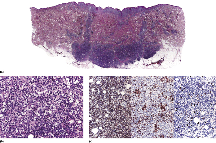
In spite of refined diagnostic criteria, better and powerful ancillary techniques, and adequate clinicopathologic correlation, some cutaneous lymphoid proliferations defy precise diagnosis and classification. Sometimes this may be due to the submission by surgeons of inadequate material (e.g., crushed specimens, superficial biopsies, or specimens that show drying artifacts – see Chapter 1), and a repeat biopsy may allow the correct diagnosis. At other times, however, an individual case cannot be classified unambiguously into a given category of cutaneous lymphoma or pseudolymphoma. I use for such cases the working term “cutaneous atypical lymphoid proliferation” (Fig. 27.1). Some of these cases show an overlap with those published in the literature as “borderline” between cutaneous lymphomas and pseudolymphomas, and are reported under various names including cutaneous lymphoid dyscrasia and clonal dermatitis, among others. It must be clearly underlined that the term “cutaneous atypical lymphoid proliferation” does not refer to cases of clear-cut cutaneous lymphoma that cannot be classified with precision. For such cases, the term “unclassifiable cutaneous (B- or T-cell) lymphoma” should be used instead, and staging investigations should be carried out.

The term “cutaneous atypical lymphoid proliferation” is mainly a histopathologic one and encompasses four main patterns:
- The first is characterized by superficial infiltrates of lymphocytes

Stay updated, free articles. Join our Telegram channel

Full access? Get Clinical Tree


