24. Treatment of the Tear-Trough Deformity
24.1 Introduction
Originally called the nasojugal groove by Loeb 1 and Duke-Elder, 2 the tear trough was so named by Flowers 3 in 1969, describing the track of the teardrop as it falls along the natural groove from the medial canthus to the cheek. Since its description, the tear trough has generated extensive discussion relating to its anatomy and the aging process that produces the tear-trough deformity. There are various causes of the tear-trough deformity, and treatment algorithms also vary, depending on the severity of the tear trough. Although relatively unremarkable in youth, the tear trough becomes progressively deeper with age and conveys an older, tired appearance.
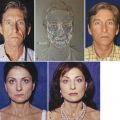
The youthful lower eyelid–cheek junction rarely displays a prominent tear trough. The aging process leads to lower eyelid fat protrusion, changes in skin texture, and distinct volume loss of the cheek fat. These factors all contribute to the prominence of the tear trough. The very nature of the thin eyelid skin, lacking subdermal fat, and the anatomically normal transition to thicker cheek skin with abundant subcutaneous fat magnify the prominence of the tear trough with age.
The prominent tear-trough deformity often conveys a tired, sad, and sleepless look that may be incongruent with the true energy level or enthusiasm of the individual. Treatment options to reverse the undesirable effects of the tear-trough deformity are thus in high demand, and surgeons have developed various interventions to diminish the appearance of the tear-trough deformity.
Unfortunately, the tear trough exists in a small and anatomically unforgiving locale. Operative intervention and alternative techniques to diminish the tear-trough deformity require thoughtful consideration of each individual’s soft tissue characteristics and the implications of treatment on both the static and dynamic appearance of the periorbital area. Although the tear-trough deformity is technically confined to the medial orbital hollow, aging often produces a progressively more bony appearance of the central and lateral orbit (i.e., loss of soft tissue with skeletonization) that may exacerbate the undesirable appearance of the tear-trough deformity by reflecting a “global hollowing” of the entire lower orbit. This transformation of the youthful, shallow-appearing orbit to an aging, hollow, deep-set orbit focuses even more attention on the tear-trough component of the aging process.
24.2 Causes
The tear-trough deformity has been attributed to various anatomical factors. Flowers mentions the descent of the cheek, a muscular gap between the orbicularis muscle and the angular head of the quadratus labii superioris muscle, hypoplasia of the suborbital malar complex, and a progressive loss of facial fat with age. 3 Some attribute the tear-trough deformity to loss of subcutaneous fat, thinning of the skin overlying the fixed orbital rim, cheek descent, and maxillary hypoplasia. 4
Others note that the tear trough overlies the muscular triangle formed by the orbicularis oculi, the levator labii superioris, and the levator alaeque nasi muscles; with advancing age, postseptal fat herniation and prezygomatic fat ptosis accentuate the defect. 5 Barton et al have described the tear-trough triad: (1) herniation of orbital fat, (2) tight attachment of the orbicularis along the arcus marginalis, and (3) malar retrusion. 6 Lambros writes that the lid–cheek junction is, in fact, stable over time, and the perception of descent is related to age-related tissue contrasts and not to actual movement. 7
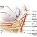
Recent studies examining the anatomy of the tear-trough deformity reveal a more precise understanding of its causes. An osteocutaneous tear-trough ligament has been described by Wong et al. 8 This ligament inserts into the skin and extends to the maxilla along the exact location of the tear trough. The tear-trough ligament becomes continuous with the orbicularis retaining ligament laterally. The “tethering effect” of the tear-trough ligament appears to be a significant factor in the development of the tear-trough deformity with aging. The groove caused by the tear-trough ligament medially and the orbicularis retaining ligamentous system laterally is the anatomical basis for the “prominent lid–cheek junction” that develops with aging. Wong et al defined the periorbital ligaments as follows: the tear-trough ligament inferomedially, which continues inferolaterally as the orbicularis retaining ligament; laterally, it becomes the lateral orbital thickening, and this continues as the periorbital septum of the upper orbit.
Other anatomical studies 5 did not visualize a distinct ligament but clearly recognized the significance of the junction of the palpebral and orbital origins of the orbicularis as closely associated with the tear-trough indentation itself, stating that “the palpebral portion of the orbicularis oculi muscle is rigidly attached to the bone, with no dissectible anatomical plane deep to the muscle.” 9 Another important distinction is that “in the suborbicularis plane,” the tear trough and the lid–cheek junction differ. Along the tear trough, it is not technically possible to dissect above the periosteum and below the muscular attachment. Along the lid–cheek junction, however, the orbicularis muscle has a ligamentous attachment to the bone by means of the orbicularis retaining ligament. These findings suggest that skin atrophy and diminished underlying subcutaneous fat are the most likely explanation for the increasing visibility of the tear trough and lid–cheek junction with age and that it is unlikely that there is age-related descent of these structures because they are fixed to the bone. This is consistent with my own clinical findings in which the thin preseptal skin with absence of subcutaneous fat is visualized cephalad to the tear trough and thick skin with ample subcutaneous fat identified below the tear trough. Additionally, the skin above the trough exhibits darker discoloration in many individuals, which seemingly “outlines” the demarcation of the tear trough. Finally, herniated fat from the medial orbital compartment will accentuate the appearance of a concavity to the adjacent tear trough. The lower lid medial fat convexity casts a shadow onto the tear trough when light is directed from above, thus emphasizing the already prominent hollow along the tear trough.
Some authors have assigned classification systems to the tear trough to provide an objective manner of evaluating the deformity. 6 , 10 Stratifying the degree of hollowing or concavity of the tear trough and the progressive loss of volume extending laterally along the lid–cheek junction is the primary focus of any analysis of the tear-trough deformity. A prominent contour “step-off” from the medial orbital rim to the cheek that extends laterally is associated with the highest grade of deformity. Some rating scales include hyperpigmentation and superficial skin rhytids that are not directly related to the depth of the trough but cast an illusion of greater depth. The recurring theme among these classifications is the progressive volume loss and concavity from medial (i.e., tear trough only) to lateral (lid–cheek junction hollowing or concavity), extending to the lateral orbit. Although categorizing the tear-trough deformity is helpful in suggesting treatment alternatives, the stratification of the deformity simply reinforces the fact that the tear- trough prominence associated with aging is simply the medial component of the aging periorbital region and that individualized treatment is necessary when considering the treatment options.
24.3 Treatment Options
A common theme for treatment of the tear-trough deformity is “leveling” of the hollow or concavity of the nasojugal groove. When the tear-trough deformity extends to the lateral orbit, volume restoration to the lid–cheek junction is indicated. To this end, some degree of filing is used, whether in the form of surgical volume restoration by repositioning tissue or direct volume addition with injection methods. Optimum success in treating the tear-trough deformity may require a combination of surgical maneuvers and injection.
Initial attempts to improve the tear trough took advantage of local fat or external implants to “level” the tear trough. Loeb 1 used local fat transposition to “fill” the concavity of the tear trough, whereas Flowers 3 used alloplastic implants and autografts to restore fullness to the tear trough. As is apparent from the pioneers who confronted the tear-trough deformity, the solution, although logical and well executed, is often less predictable and effective than desired. Incomplete resolution of the tear trough (i.e., failure to level the concavity) and visible surface irregularities may be encountered. Recent anatomical studies more clearly define the tear-trough deformity, and yet, despite greater anatomical understanding, surgical endeavors to treat the tear-trough deformity are still influenced by the unforgiving nature of the lower eyelid, manifested by often unpredictable consequences of scar contracture, eyelid malposition, static and dynamic contour irregularities, and inadequate tear-trough correction.
Injection techniques have gained popularity secondary to their perceived ease, minimal downtime, and the general allure of a nonoperative treatment option. Modern-day “off-the-shelf” fillers such as the hyaluronic acid products garner the most use because of their safety profile and reversibility. Autologous fat injection also remains a viable option in experienced hands, although long-term static and dynamic (animation) deformities may be difficult to resolve. The undesirable visibility of injected material and the limited success of injection techniques (only providing minor degrees of change of the tear-trough deformity) dampen the unbridled enthusiasm for injectable fillers.
Regardless of the chosen approach for treatment of the tear-trough deformity, precise anatomical analysis, technical skill, and individualized treatment application are essential to an aesthetically pleasing correction.
24.3.1 Injections
Injectable filler materials that provide volume restoration are the most common nonsurgical option for correction of the tear-trough deformity. Autologous fat and hyaluronic acid fillers have become the treatment mainstays for many surgeons. Injections are most successful in individuals with minor tear-trough deformities, typically patients with good skin tone and minimal medial fat protrusion. 7 , 10 , 11 , 12 , 13
The tear trough or nasojugal groove is characterized by the junction of thin eyelid skin and thick cheek skin. Injectable filler techniques can be effective in leveling the tear-trough deformity; however, this requires precise execution and careful anatomical placement of the filler material. 14 The best candidates for nonsurgical treatment are those with thicker, smoother skin, a clearly defined hollow, and minimal skin hyperpigmentation, as pigment changes are not improved with fillers. 4 Furthermore, patients with significant medial orbital fat protrusion will likely have limited success with fillers.
24.3.2 Hyaluronic Acid Fillers
The best plane for injection of filler material is debatable. Use of the superficial injection plane in the deep dermal or subdermal space essentially is an attempt to thicken the anterior lamella and provide surface support to the thin skin of the tear-trough area. 15 The superficial approach requires great patience and skill to avoid irregularities and is adopted less frequently than the deep plane technique. Injection of fillers deep to the orbicularis oculi muscle is the technique preferred by most surgeons, as the risk of surface nodules and irregularities is reduced. Hyaluronic acid fillers may be placed into the orbicularis muscle or deep to the muscle, whereas percutaneous injected fat is best placed deep to the muscle.
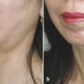
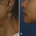
I limit the indication for hyaluronic acid filler treatment to minor tear-trough deformities. Patients who exhibit considerable herniated orbital fat and global hollowing of the entire periorbital region are offered surgical alternatives. My preferred technique for injection of the tear-trough deformity with hyaluronic acid fillers is in the suborbicularis space, although in patients with thicker skin texture, injection may also be placed into the orbicularis muscle. Injections proceed from the midpupillary line of the midorbit and are directed medially. The injection is placed just below the actual concavity of the tear trough, consistent with anatomical studies showing that placement of filler in the orbicularis muscle and suborbicularis regions is preferred to more superficial injections. A blunt, side-port 27-gauge needle is used in the deep injection plane, and a 32-gauge needle is used for injection more superficially into the muscle. Dermal injections are rarely indicated and are performed with great caution, injecting miniscule amounts of filler materials. All hyaluronic acid filler injections into the tear trough involve multiple passes with gentle plunger pressure and crossing patterns to avoid linear deformities from unidirectional injection. The injected filler volume is typically between 0.2 and 0.5 mL per side. Postinjection massage of the treated area is limited to avoid further trauma, and application of a cold compress is suggested for 12 hours posttreatment.
Related posts:
 6. Facial Sculpting and Facial Slimming with Neurotoxins
 20. Comparison of Midface Rejuvenation Techniques
 22. Lower Eyelid Blepharoplasty
 5. Energy-Based Treatments for Facial Aging
 9. Male versus Female Facelift Surgery. Is There a Difference?
 12. Primary Superficial Musculoaponeurotic System (SMAS) Facelift and Neck Lift
Stay updated, free articles. Join our Telegram channel

Full access? Get Clinical Tree


