20. Comparison of Midface Rejuvenation Techniques
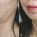
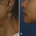
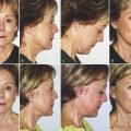
The “midface” has received significant attention in the plastic surgery literature. In this chapter, we review the relevant anatomy of the midface and the various surgical procedures that have been used to improve the midfacial area. A consistent characteristic reported in the literature is that procedures directed specifically to the midface are applicable to only a very few patients. When midfacial lifts are applied to older patients, additional procedures are required to obtain an acceptable result. A high–superficial musculoaponeurotic system (SMAS) facelift with possible dermal fillers or fat grafting is the procedure of choice to correct the problems associated with the aging face and provides for a harmonious and comprehensive treatment. High-SMAS deep plane support allows for ancillary procedures that enhance the patient’s pleasing appearance.
Facelifting has evolved since its skin excision and tightening origins in the early 20th century. Deep plane techniques by Skoog 1 and the description of the SMAS by Mitz and Peyronie 2 conveyed a new era of facelifting that allowed for significant improvement in the cheeks and neck. 3 , 4 , 5 , 6 , 7 , 8 , 9 , 10 , 11 , 12 Limitations in these procedures have led some authors to develop techniques to address the midface specifically. 13 Many different approaches have improved the changes observed with aging of the midface. These are summarized in Table 20.1. The purpose of our review is to compare the various modalities of midfacelifting.
20.1 Relevant Anatomy
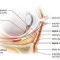
The superior border of the midface has been defined as the inferior orbital rim. The inferior border is at the level of the oral commissure, the medial border is the nasolabial crease, and the lateral border is the anterior border of the masseter muscle. 14 A key anatomical feature of the midface is the malar fat pad. 15 Malar fat pad ptosis is a significant component of the changes associated with the aging midface. There is also a loss of subcutaneous tissues over the infraorbital rim. Laxity of the orbital septum and ptosis of the malar fat pad lead to the double convexity seen on a lateral view of the patient and an increased lower eyelid-to-cheek distance. Ptosis of the malar fat pad also produces prominence of the nasolabial folds.
20.2 Coronal Subperiosteal Facelift
An early attempt to specifically address the midface was development of the subperiosteal facelift performed via a coronal approach. 6 , 8 , 11 , 12 The technique was pioneered by Tessier 11 and popularized by Psillakis et al. 8 The procedure involves a subperiosteal dissection of the face via a coronal incision, similar to the approach used to repair a facial fracture or perform midfacial osteotomies. A gingivobuccal sulcus incision may be added to free the lower midface completely. Sutures are then placed in the periosteum of the origin of the zygomaticus major muscle, and the periosteum is suspended and fixed to the temporalis fascia. The periosteal suspension provides strong elevation of the midface.
The subperiosteal approach has several advantages. The soft tissues including the facial nerves are all kept superficial to the plane of dissection. This is advantageous for the novice surgeon because there is less potential injury to the zygomatic and buccal branches of the facial nerve than with deep plane and SMAS procedures. The blood supply to the skin flaps is also maximally maintained, and there are no long skin flaps with the potential for skin-flap necrosis.
This approach has some significant disadvantages associated with it as well. The subperiosteal approach repositions the origins of the zygomatic musculature and may result in excessive horizontal width to the midface. More facial edema may be present postoperatively than with other approaches. Most surgeons perform the subperiosteal dissection over the anterior portion of the zygomatic arch, which places some risk to the temporal branch of the facial nerve. Finally, if there is any significant lower facial laxity, another procedure, such as a lower facelift, must be performed to obtain an acceptable result.
20.3 Transtemporal Endoscopic Midfacelift
The endoscope began to be applied for plastic surgery procedures beginning in the mid-1990s. Initial efforts for facial rejuvenation were focused on elevation of the brow, but subsequent efforts applied the use of the endoscope for midface elevation combined frequently with browlifting procedures. The endoscope enables the use of smaller incisions and provides excellent visualization and magnification of the tissues. Most surgeons have used the subperiosteal plane, 16 , 17 , 18 , 19 but others have used a supraperiosteal plane. 20 A multiplane approach has also been applied to the midface. 21
Patients who are candidates for this procedure tend to be younger patients with isolated midfacial aging and early jowls, patients who are not ready for a more extensive surgical procedure, and those patients who will not accept a preauricular scar. Patients in whom there is more significant laxity of the skin will require an additional lower face rhytidectomy that may involve elevation of an SMAS flap, thus limiting the utility of the transtemporal endoscopic midfacelift approach.
Most surgeons use three incisions about 2 cm long posterior to the hairline. Additionally, a longer incision is used posterior to the temporal hairline. The initial dissection is in the subgaleal plane. As the dissection proceeds more inferiorly, several different planes have been chosen, including staying directly on top of the superficial layer of deep temporal fascia, dissecting just underneath the superficial layer of deep temporal fascia, and going over the top of the deep layer of deep temporal fascia and elevating the intermediate fat pad and superficial layer of deep temporal fascia. Subperiosteal dissection is then continued over the zygomatic arch and zygoma. A gingivobuccal sulcus incision may be used to obtain adequate release of the lower face. Sutures are then placed into the malar fat pad or into the area of the origin of the zygomaticus major muscle. The midfacial tissues are suspended superiorly and posteriorly and then fixated to the temporalis fascia or held in place with bone screws. Alternatively, an Endotine device may be used to suspend the midfacial tissues. The superior end of the Endotine device is then sutured to the temporalis fascia. 17
The reported advantages of the endoscopic approach are hidden, small incisions that allow clear view of the anatomy of the region. The tissues of the midface can be released and then repositioned as a unit.
Our review of the published series shows a clear augmentation of the malar area; however, the patients all seem to require ancillary procedures to achieve a quality result. This is especially true in the lower face where a “lower facelift” is required to obtain an effect on the lower face, jowls, and neck. The transtemporal endoscopic midfacelift does not appear to produce any effect on the corners of the mouth, and there is a limited effect on the nasolabial fold.
20.4 Transblepharoplasty Midfacelift
Hester and colleagues initiated the era of elevation of the midface using a transblepharoplasty approach. 22 Subsequent surgeons have also used the transblepharoplasty approach using either a subperiosteal 23 , 24 or a preperiosteal approach. 25 Candidates for the procedure are also typically younger patients with midfacial aging.
The procedures typically involve a subciliary incision. Dissection is performed inferiorly in the subcutaneous plane, or there is elevation of a skin–muscle flap after preserving a strip of pretarsal muscle. In the subperiosteal procedures, the periosteum is opened after leaving an intact cuff of periosteum, and subperiosteal dissection proceeds inferiorly, taking care not to injure the infraorbital nerve. The periosteum may or may not be released at the caudal aspect of the dissection. The preperiosteal dissection extends inferiorly for a distance of about 4 cm from the orbital rim. The cheek soft tissues are then suspended either to the lateral orbital rim or to the temporal fascia. Adjunctive procedures may include the performance of a canthopexy or canthoplasty, liposuction of a cheek bulge in the lateral orbital rim, and elevation of the skin from the subcutaneous tissues from a temporal incision to prevent bunching of the skin in the lateral canthal area.
The transblepharoplasty approach has a significant rate of complications after the procedure. The issues have included lateral canthal malposition, scleral show, problems with dry eyes, and injury to the infraorbital nerve. Perhaps most important is the need for adjunctive procedures to achieve an acceptable result, including lower facelifting, laser treatments, and chemical peels.
Related posts:
 6. Facial Sculpting and Facial Slimming with Neurotoxins
 22. Lower Eyelid Blepharoplasty
 17. Secondary Facelifting
 5. Energy-Based Treatments for Facial Aging
 9. Male versus Female Facelift Surgery. Is There a Difference?
 12. Primary Superficial Musculoaponeurotic System (SMAS) Facelift and Neck Lift
Stay updated, free articles. Join our Telegram channel

Full access? Get Clinical Tree


