Skin Signs of Systemic Cancers 
Mucocutaneous Signs of Systemic Cancers
ICD-9: 199.0  ICD-10: M8000/6
 Mucocutaneous findings may suggest systemic cancers in several ways:
 Associations of heritable mucocutaneous disorders with systemic cancers.
 By action at a distance, i.e., paraneoplastic syndromes.
 Or spread of cancer to the skin or mucosal sites by direct, lymphatic, or hematogenous extension (cutaneous metastasis).
Metastatic Cancers
Persistent Tumor. Lymphatic extension, hematogenous spread.
Direct Extension. Paget disease, extramammary Paget disease.
Lymphomas with secondary skin involvement (Section 21).
Heritable Disorders
Cowden Syndrome
Peutz–Jeghers Syndrome
Neurofibromatosis (p. 405).
Tuberous sclerosis (p. 402).
Multiple endocrine neoplasia (types 1 and 2b).
Paraneoplastic Syndromes
Acanthosis nigricans, malignant, tripe palms.
Acquired ichthyosis.
Bazex syndrome.
Carcinoid syndrome.
Dermatomyositis (p. 328).
Ectopic ACTH syndrome.
Erythema gyratum repens.
Gardner syndrome.
Glucagonoma syndrome.
Hypertrichosis lanuginosa.
Muir–Torre syndrome.
Palmar keratoses.
Paraneoplastic pemphigus.
Pruritus.
Pyoderma gangrenosum (p. 116).
Sweet syndrome (p. 120).
Vasculitis (p. 356).
Metastatic Cancer to the Skin* ICD-9: 199.0  ICD-10: M8000/6
 Metastatic cancer to the skin is characterized by solitary or multiple dermal or subcutaneous nodules, occurring as metastatic cells from a distant noncontiguous primary malignant neoplasm.
 They are transported to and deposited in the skin or subcutaneous tissue by one of the following routes:
 Lymphatic routes.
 Hematogenous spread.
 Contiguous spread across the peritoneal cavity or other tissues.
 Skin lesions nodule (Figs. 19-1 and 19-2), raised plaque, thickened fibrotic area. First detected when <5 mm. Fibrotic area may resemble morphea; occurring on scalp, may produce alopecia. Initially, epidermis is intact, stretched over nodule; in time, surface may become ulcerated (Fig. 19-3) or hyperkeratotic. May appear inflammatory, i.e., pink to red or hemorrhagic. Firm to indurated. May be solitary, few, or multiple. May acquire considerable size and may be mistaken for a primary skin cancer (Fig. 19-3).
*For metastatic nonmelanoma skin cancers and melanoma, see Sections 11 and 12.
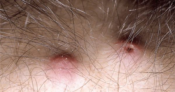
Figure 19-1. Metastatic cancer to the skin: bronchogenic cancer Dermal nodules on the scalp of a patient undergoing chemotherapy for metastatic lung cancer; the nodules were only apparent following loss of hair during chemotherapy. The nodule on the left is asymptomatic, erythematous, but noninflamed. The nodule on the right has a central depression marking a punch biopsy site.
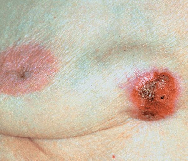
Figure 19-2. Metastatic cancer to the skin Breast cancer: Large nodule on breast in a 40-year-old woman with breast cancer, present for 4 months.
Figure 19-3. Metastatic cancer to the skin Adenocarcinoma of the GI tract. This fungating mass was just the tip of the iceberg: a much larger mass was in the subcutis.
Special Patterns of Cutaneous Involvement
Breast
Inflammatory metastatic carcinoma (carcinoma erysipelatodes): erythematous patch or plaque with an active spreading border (Fig. 19-4). Most often with breast cancer that may spread within lymphatics to the skin of involved breast, resulting in inflammatory plaques resembling erysipelas (hence, the designation carcinoma erysipelatodes). Occurs with other cancers as well [pancreas, parotid, tonsils, colon, stomach, rectum, melanoma, pelvic organs, ovary (Fig. 19-5), uterus, prostate, lung, mesothelioma (Fig. 19-6)].
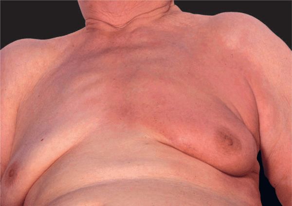
Figure 19-4. Metastatic cancer of the skin: inflammatory breast cancer (carcinoma erysipelatodes) A large erythematous and only minimally indurated lesion covering the entire breast and presternal region; the lesion is red and sharply defined and thus looks like erysipelas. There was a 2 × 2 cm lump in the breast upon palpation.
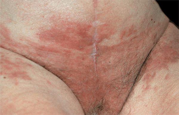
Figure 19-5. Metastatic ovarian cancer Manifesting as carcinoma erysipelatodes on the lower abdomen and inguinal region. Workup disclosed ovarian cancer with peritoneal carcinomatosis.
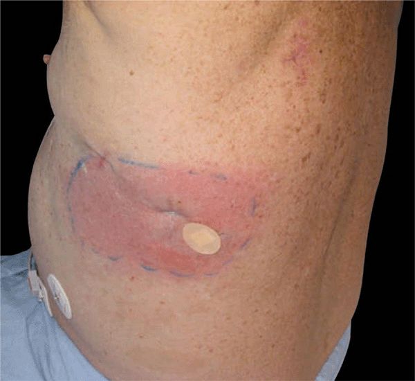
Figure 19-6. Mesothelioma An indurated erythematous patch on the lateral chest represents carcinoma erysipleatodes from mesothelioma.
Telangiectatic metastatic carcinoma (carcinoma telangiectaticum): breast cancer appearing as pinpoint telangiectases with dilated capillaries within carcinoma erysipelatodes. Violaceous papules or papulovesicles resembling lymph-angioma circumscriptum.
En cuirasse metastatic carcinoma: diffuse morphea-like induration of skin (Fig. 19-7). Usually local extension of breast cancer occurring in breast and presternal region. Sclerodermoid plaque may encase chest and resembles a metal breastplate of a cuirassier. Also occurs with primary of lung, GI tract, kidney.
Multiple smooth nodules on scalp: prostate adenocarcinoma, lung cancer, breast cancer (Fig. 19-1).
Alopecia neoplastica: On scalp, areas of hair loss resembling alopecia areata; well-demarcated, red-pink, smooth surface, flat.
Large Intestine. Often presents on skin of abdomen or perineal regions; also, scalp or face. Most originate in rectum. May present with metastatic inflammatory carcinoma (like carcinoma erysipelatodes) of inguinal region, supraclavicular area, or face and neck. Less commonly, sessile or pedunculated nodules on buttocks, grouped vascular nodules of groin or scrotum, or facial tumor. Rarely, cutaneous fistula after appendectomy or resembling hidradenitis suppurativa.
Lung Carcinoma. May produce a large number of metastatic nodules in a short period. Most commonly, reddish nodule(s) on scalp (Fig. 19-1). Trunk: symmetric; along direction of intercostal vessels, may be zosteriform; in scar (thoracotomy site or needle aspiration tract).
Hypernephroma. Can produce solitary lesion; also widespread. Usually appear vascular, ± pulsatile, ± pedunculated; can resemble pyogenic granuloma. Most common on head (scalp) and neck; also trunk and extremities.
Carcinoma of Bladder, Ovary. Can spread contiguously to abdominal and inguinal skin similarly to breast cancer, as described above, and look like erysipelas (Fig. 19-5).
Miscellaneous Patterns. With dilation of lymphatics and superficial hemorrhage, may resemble lymphangioma. With lymph stasis and dermal edema, resembles pigskin or orange peel.

Figure 19-7. Metastatic breast cancer: cancer en cuirasse Both breasts are hard upon palpation—like an armor plate. There are multiple small and large, ulcerated nodules and there is a background of erysipelas-like erythema (carcinoma erysipelatodes).
Mammary Paget Disease ICD-9: 174.0  ICD-10: C50.01
 Mammary Paget disease (MPD) is a malignant neoplasm that unilaterally involves the nipple or areola and simulates a chronic eczematous dermatitis.
 It represents contiguous spread of underlying intraductal carcinoma of the breast (1–4% of breast cancers).
 Usually occurring in females (>50 years); there are rare examples in males.
 Onset is insidious over several months or years. May be asymptomatic or there may be pruritus, pain, burning, discharge, bleeding, ulceration, and nipple invagination.
 Skin lesion presents as red, scaling plaque, rather sharply marginated, oval with irregular borders. When scale is removed, the surface is moist and oozing (Fig. 19-8). Lesions range in size from 0.3 to 15 cm (Fig. 19-9). In early stages, there is no induration of the plaque; later, induration and infiltration develop and nodules may be palpated in breast. At initial, there is flattening or retraction of the nipple presentation, an underlying breast mass is palpable in fewer than one-half of patients. May be bilateral. Lymph node metastases occur more often when MPD is associated with an underlying palpable mass.
 Differential diagnosis includes eczematous dermatitis, psoriasis, benign ductal papilloma, nipple-areola retention hyperkeratosis, impetigo, SCC in situ, familial pemphigus.
 Eczematous dermatitis of the nipples is usually bilateral; it is without any induration and responds rapidly to topical glucocorticoids. Nevertheless, be suspicious of Paget disease if “eczema” persists for >3 weeks. Diagnosis verified by biopsy showing neoplastic cells in epidermis following a pathognomonic pattern of spread. Define underlying intraductal carcinoma by mammography.
 Management consists of surgery, radiotherapy, and/or chemotherapy as in any other breast carcinomas. Lymph node dissection if regional nodes are palpable. Prognosis varies. When breast mass is not palpable, 92% of patients survive 5 years after excision; 82% survive 10 years. When breast mass is palpable, 38% survive 5 years; 22% survive 10 years. Prognosis worse when there is lymphadenopathy.
Related posts:

Stay updated, free articles. Join our Telegram channel

Full access? Get Clinical Tree


