16 Bilateral Septal Mucosal Flaps in Septal Perforations
Summary
Septal perforations repair is a very challenging and sometimes frustrating surgery for the nose surgeon; we describe a technique based on an extended lateral dissection of the mucosa. For this, we can use either the external approach or the endonasal endoscopic approach. The lateral dissection of the flaps extends superiorly under the nasal bones and lateral superior cartilages and inferiorly under the inferior turbinate. For flaps closure, a transfixing suture is preferred to avoid tension and tearing of the flaps.
16.1 Introduction
As a rhinoplasty surgeon, I frequently have to deal with septal perforations in two different situations. First, there are patients with symptomatic perforations due to previous surgery or other previous causes, and second, there are patients in whom the perforation occurs during surgery when the mucosa is bilaterally damaged, which subsequently needs mucosal fixation. It occurs especially in revision surgery.
There are many options to treat nasoseptal perforations, from prosthetics 1 to a number of different types of flaps with or without interposition of tissue. The literature shows the results often contradictory and rarely statistically significant. 2 , 3
To repair these perforations, I use a modified technique for hump removal with mucosal preservation. This can be done with an external or endoscopic approach. Both of these techniques need a good dissection of the mucosa and to suture without tension of the flaps.
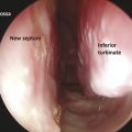
For this technique, it is very important to know the anatomy of the septum. Review s. Kap. to get greater insights about blood supply of the nose and nasal septum. Bilateral septal mucosal flaps are dissected in a submucopericodrial-mucopieriosteal plane from anterior to posterior and from superior to inferior, thus preserving the vascular supply of the posterior and superior portion of the septum.
The septal artery network comes from the septal artery that runs over the rim of the posterior choana after the division of the sphenopalatine artery at the level of the sphenopalatine foramen. These arteries run from posterior to anterior so they allow for a good dissection of the bilateral flaps (Fig. 16‑1a, b).
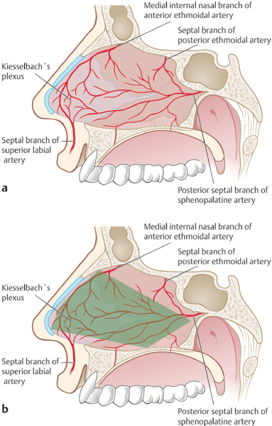
The system of the internal carotid artery with one of its branches, the ophthalmic artery, divides into the anterior and posterior ethmoidal arteries, which also play a role in the vascularization of this flap.
16.2 Indications
The advancement of local flaps alone or combined with interposition of grafts techniques is suitable for small to moderate symptomatic septal perforations with good superior and inferior margins. 4
The main symptoms of nasal septal perforation that we find are crusting, epistaxis, and nasal obstruction, as in the literature. In our patients, these symptoms are worse due to the altitude (2,800 m) and lack of humidity.
Some patients may feel some relief with conservative treatment such as humidification, moisturizing ointments, and nasal saline irrigations; however, these measures have a limited and temporary effect. 5
Patients with active cocaine abuse, topical nasal vasoconstriction spray overuse (oxymetazoline), systemic disease (granulomatosis with polyangiitis), those who play contact sports, and those who have other similar conditions that impair a good vascular supply are not good candidates for this technique.
16.3 Surgical Technique
The bilateral mucosa flaps technique is based on the septal mucoperichondrial flap, which is insufficient for most of the nasoseptal perforations, and needs to be completed with lateral extension mucoperiosteal flaps in the floor and under the nasal bones. The use of endoscopes is mandatory for the dissection.
Lee et al 6 describe a similar endoscopic technique, but they dissect just one side and use temporalis fascia between the flaps.
16.3.1 Instrumentation
The instrumentation used for this technique is the same as for rhinoplasty when the open approach is used. If we choose the endoscopic approach, a 0-degree endoscope is the standard for mucosal dissection. A 30- or 45-degree endoscope is useful for the lateral extension of the dissection under the nasal bones and in the floor of the nose under the inferior turbinate. For the mucosa edges, a polyglactin (Vicryl) 5–0 is used for suturing.
16.3.2 Technique
Step 1
Local and General Anesthesia
All the patients are operated on under general anesthesia to obtain complete amnesia, analgesia, and sedation. The local anesthesia protocol that we use is as follows:
Lidocaine 2% in combination with epinephrine 1:200,000
Oxymetazoline 0.050% embedded in two neurosurgeon pledges
A 27-gauge needle with a 5-cc syringe is used to infiltrate the local anesthetic in the subperichondrial-subperiosteal plane where there is cartilage or bone, and between both mucosa where there is no cartilage or bone. This usually happens around the perforation, especially in the case of postseptoplasty patients. In these patients in whom the loss of cartilage and bone is bigger than the perforation, it is very useful to put the local anesthetic in the right plane to help us with the dissection.
We begin the infiltration in the posterior part of the nasal septum and then we continue with small amounts of local anesthetic anteriorly. This helps us to avoid bleeding in the area of injection.
Finally, we inject the floor of both cavities, under the nasal bones and superior lateral cartilages. Cottonoids are placed in each nasal cavity. At this time, the vibrissae are cut with a number 15 blade for maximum visualization during surgery.
Related posts:

Stay updated, free articles. Join our Telegram channel

Full access? Get Clinical Tree


