13 Anterior Ethmoidal Artery Septal Flap
Summary
Nasal septal perforation (NSP) is a defect of the nasal cartilage and/or nasal bone septum with an approximate prevalence of 1% in an adult population, although it has probably been underestimated because many patients remain asymptomatic. There are different surgical treatment options to close the perforation. The authors discuss the anterior ethmoidal artery septal flap. This flap is mainly indicated in symptomatic patients with anterior septal perforation. A unilateral mucosal flap with a large and flexible pedicle should be harvested to bring a suitable blood supply to the flap. The extension to the inferior meatus may create additional mucosal flap, allowing advancement of the flap without tension.
13.1 Surgical Anatomy
Numerous flap designs have been described in the literature to close septal perforations. Advancement or rotation of mucoperichondrial/mucoperiosteal tissue from the nasal septum or nasal floor has been widely utilized.
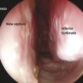
This study focused on the use of a monolateral mucosal septal flap pedicled on septal branches of the anterior ethmoidal artery (AEA), extended to the nasal floor and the inferior meatus.
Knowledge about the vascularization of the nasal septum is crucial if aiming to preserve its branches during the incision of mucoperichondrium and mucoperiosteum to harvest the septal flap.
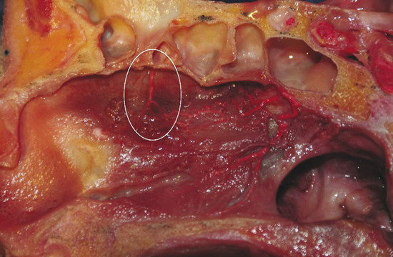
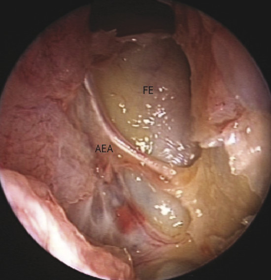
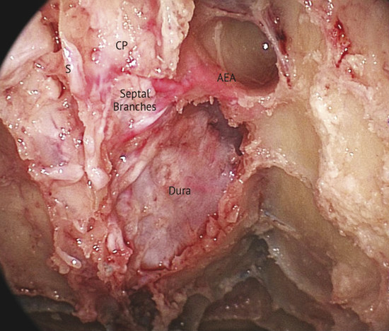
Blood supply of the nasal septum occurs through septal branches of the sphenopalatine artery. Here it anastomoses with branches of the palatine and labial arteries and septal branches of the anterior and posterior ethmoidal arteries; they are easily recognizable in the cranial portion of the septum area (Fig. 13‑1). An anatomical study on the arterial pattern of the nasal septum, traced by microdissection, demonstrated that AEAs were present in all cases, but the posterior ethmoidal arteries in some cases were absent. 1 These arteries, with the middle septal branch of the sphenopalatine artery and the superior labial branch of the facial artery, mainly contribute to the anastomotic triangle of the anterior septum. Only the posterosuperior area is vascularized by the posterior ethmoidal artery branches. 2 The ethmoidal artery originates from the terminal segment of the ophthalmic artery in the orbit cavity, a collateral branch of the internal carotid artery, and passes between the superior oblique and medial rectus muscle. The AEA then reaches the frontoethmoidal suture through the anterior ethmoidal foramen and enters into the anterior ethmoidal canal along with the anterior ethmoidal nerves. The artery crosses the ethmoid roof diagonally from posterolateral to anteromedial (Fig. 13‑2). AEA then divides at the lateral part of the cribriform plate of the ethmoid, giving off two or three branches to the mucosa of the cranial portion of the septum (Fig. 13‑3). Finally, they reach the olfactory cleft and supply terminal branches to the olfactory bulb and the meninges.
One must consider that the septal branches of the AEA are at the same level or just posterior to the septal projection of the axilla of the middle turbinate, as confirmed by a recent anatomical analysis, which showed that the average distance was 7.35 mm (range: 5.5–8.7 mm) and never superior to 1 cm. 3
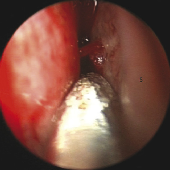
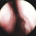
Bleeding can be seen originating from a septal branch of the AEA, showing a close anatomic relation with the axilla of the middle turbinate (Fig. 13‑4). It is extremely important to consider this anatomical relation during harvesting the flap.
13.2 Relevant Analytical Factors
The size and the location of the perforation play a large part in the decision-making process in performing surgery and in choosing the most suitable technique. Symptomatology is essential in determining whether a perforation should be repaired. If the perforation is in the posterior part of the septum and asymptomatic, repair is rarely necessary.
All perforations of our series were located in the anterior part of the septum and the mean size of the septal perforation was 15 mm (range: 10–25 mm).
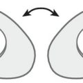
Based on the anatomical criteria of the nasal septum vascularization, characterized by the constant presence of the septal branches of the AEA, we harvested the monolateral mucosal flap with a large and flexible pedicle to bring a suitable blood supply to the flap. The extension to the inferior meatus creates a larger mucosal flap, allowing advancement of the flap without tension. 4
These two aspects, the flap designed without tension and the preservation of the flap’s vascular supply, are the main factors contributing to a high rate of closure.
With our technique the normal respiratory mucosa of the nose is used for reconstructing the anatomy and physiology of the nasal septum.
This septal flap is not useful for posterior perforations because the flap cannot be extended to the posterior part of the septum, where the mucoperiosteum is elevated.
Regarding the necessity for bilateral flap provision, monolateral flap coverage was advocated by some authors, as it limits the donor area to one side of the nose, and thus preserves more nasal respiratory mucosa while achieving favorable closure rates. 5 , 6 , 7
Unlike previous studies, a recent case series suggested that NSP could be successfully repaired without using interposing grafts. 8 Even with incorporating grafts, surgical outcomes can vary according to the types of flap utilized for the repair, so the success related to interpositional graft actually is not considered statistically significant. 9
Furthermore, because no cartilage graft is harvested, it also abolishes donor site morbidity. The choice to use a monolateral flap and avoid interposition of any graft may help reduce operating time.
At the end of the procedure, we recommend the use of Silastic sheets to avoid postoperative scarring.
The endoscopic endonasal approach, in our opinion, is adequate for surgical exposition of anterior septal perforations, flap dissections, and suturing. The use of nasal endoscope allows superior precision in all surgical steps by ensuring excellent access to the operating sites. A critical factor is having a good view of the surgical field and the anesthesiologist’s contribution that are essential in achieving an effective hemostasis.
Related posts:

Stay updated, free articles. Join our Telegram channel

Full access? Get Clinical Tree


