11. Endoscopic Browlift
11.1 Introduction
Forehead aging is a common aesthetic concern in aging patients. It reflects or communicates fatigue, anger, and sadness in affected individuals. Commonly encountered signs of forehead aging include brow deflation and ptosis with lowering of the medial and lateral hair-bearing brow and development of forehead and glabellar wrinkles. Browlift is a surgical procedure that addresses the drooping brow and rejuvenates the forehead.
Multiple techniques and approaches have been described for browlifting. They can generally be divided into open and endoscopic techniques. Open techniques include coronal, temporal, midforehead, anterior hairline, and transpalpebral approaches. 1 , 2 , 3 , 4 , 5 , 6 , 7 , 8 , 9 , 10 These techniques vary with respect to the site of incision, plane and method of dissection, and type of fixation. Generally, most of these techniques, both open and endoscopic, involve mobilization, repositioning, and fixation of the forehead and alteration of the forehead muscles.
In most surgical specialties, technology has created a trend toward less invasive techniques. Plastic surgery and browlift are no exception. Endoscopy has been used by gynecologists since the 1970s for diagnostic and therapeutic purposes. In the mid-1980s, endoscopy was introduced into general surgery, and by 1990 laparoscopic procedures became standard of care in general surgery. The first endoscopic browlift was performed at the University of Alabama in 1992. The endoscopic approach was developed as an alternative to the open techniques in selected patients to avoid long transverse incisions and their potential complications. Also, it can improve intraoperative magnification and visualization of small anatomical structures and allows more accurate dissection. 11 Over the last two decades, endoscopic techniques have evolved significantly, instruments have improved, and relevant anatomy has been better defined. Currently, endoscopic browlift is one of the most commonly performed endoscopic procedures in plastic surgery.
11.2 Authors’ Personal Experience
Consensus on browlifting has been hard to come by. After initially being a “blackballed” procedure in the United States, the open browlift gradually became an accepted part of the plastic surgeon’s armamentarium (personal communication, Bruce Connell, 1997).
Over time, surgeons and patients alike became disenchanted with the drawbacks of coronal browlifting. Some of these were technique and surgeon dependent, whereas others were inherent parts of the nature of the procedure. 12 , 13 With the introduction of the endoscopic browlift, a renewed enthusiasm for the benefits of brow–forehead rejuvenation emerged. As with all things, this enthusiasm “at times runneth over,” leading to a battle for whose and which technique would produce the highest and most lasting elevation. Lost in the “race to the top” was the aesthetic and beauty sense that would define the success of the procedure. As a result, practices have divided into two camps.
One faction has dismissed and discarded the browlift entirely, citing complications and unnatural creations. 12 , 13 A second group has sought the aesthetics of natural beauty. While this trail has been evolving, a variety of approaches that may be effective in unique situations have been developed. 1 , 2 , 3 , 4 , 5 , 6 , 7 , 8 , 9 , 10 Also, refinements in the endoscopic browlift have emerged to become the mainstay for those who decline to “throw out the baby with the bathwater.” 11 , 14 , 15 , 16
Over time, in the practitioners’ browlift experience, as well as in the literature, there has been an increased appreciation for the aesthetics of youth and natural beauty, as well as individual variation. To try to create an elegant arch on someone with a naturally low, straight brow works no better than to cut and paste a large broad smile onto a small thin face; the mismatch and imbalance offend the eye.
Nevertheless, there are certain consistent, cross-cultural, transcontinental desirable features, whether in South America or Scandinavia. Those include smooth, full, soft tissue drape over the lateral orbital rim; no fold past the corner of the eye; an arch (if present) that peaks between the medial two-thirds and lateral one-third of the brow; and the lack of vertical glabellar furrows medially. In addition, minimalization of static transverse forehead lines is desirable.
Given these features, we have developed a standardized method for evaluating the brow–forehead complex using this methodical approach. A customized aesthetic improvement can be offered to each individual patient.
11.3 Pertinent Anatomy
11.3.1 Scalp and Frontalis Muscle
Scalp layers include the skin, subcutaneous tissue and fat, aponeurosis (galea aponeurotica), a loose areolar connective tissue plane, and the periosteum. As the scalp transitions into the forehead, the frontalis muscle is found between the subcutaneous tissue and galea. The frontalis muscle has no bony attachments; its fibers blend with procerus, corrugator, and orbicularis muscles. Its primary function is to lift the eyebrows.
11.3.2 Temporal Area
From deep to most superficial, the temporal layers include periosteum, temporalis muscle, temporalis fascia, a loose areolar plane, temporoparietal fascia, and subcutaneous tissue and skin. The temporal crest marks the transition from the temporoparietal fascia to the periosteum of the frontal region.
11.3.3 Vascular Anatomy
Supraorbital vessels arise from the supraorbital foramina bilaterally. These arteries, along with the superficial temporal and the occipital arteries, contribute to the rich blood supply to the scalp. In addition, the supratrochlear arteries arise medially to the supraorbital vessels and contribute to the perfusion of the central forehead.
11.3.4 Sentinel Vein
Zygomaticotemporal veins are communicating veins between the deep and superficial systems. Usually, there is one large medial vein, referred to as the sentinel vein, and a smaller lateral vein. It is important to mark the sentinel vein preoperatively. Some surgeons advocate preserving this vein in endoscopic dissections; others recommend its cauterization.
11.3.5 Neurological Anatomy
The forehead sensory innervation is supplied by the supraorbital and supratrochlear nerves. These nerves exist in neurovascular bundles with their corresponding arteries. Usually, a single dominant sensory nerve is present. The supraorbital nerve exits through the notch at around 2.7 cm from midline; the supratrochlear nerve is usually located at 1.5 to 2 cm from midline. These nerves are commonly seen in endoscopic browlift and usually lie within or superficial to the depressor supercilii and corrugator muscles. Another nerve to be cautious of during this dissection is the temporal branch of the facial nerve. The lowest branch of this nerve passes approximately 1 cm posterosuperior to the sentinel vein.
11.3.6 Glabellar Muscles
During endoscopic browlift, the corrugator supercilii is usually the first muscle to be encountered. The anatomical description of the corrugator muscle has evolved over the last decade, and the dimensions of the muscle are more extensive than once thought. Cadaveric studies describe the muscle to be around 3 cm from the nasion medially and 14 cm laterally. 17 Medial to the corrugator, we encounter the depressor supercilii muscle, and more superficially lies the procerus muscle. It is important to know that the supratrochlear nerve runs within or superficial to the corrugator muscle. 18
11.4 Advantages and Disadvantages of Endoscopic Browlift 18
11.4.1 Advantages
Shorter and less visible scars
Intraoperative magnification of structures
Exposure of periorbital adhesions, hence better release
Reduced risk of alopecia and scalp sensory changes
Reduced operative time
Less tissue disruption, hence quicker recovery
Avoids reliance on “crutch” of skin tension and resultant negative consequences
11.4.2 Disadvantages
Equipment cost
Technology-dependent nature of procedure; failure can prevent completion
Operator learning curve
Need for additional fixation
Reliance on precise deep tissue alteration (in the absence of skin removal)
11.5 Preoperative Assessment
The importance of a thorough patient interview during the initial consultation cannot be overemphasized. It is during this encounter that the surgeon and the patient start establishing a personal relationship that creates a comfortable environment, which is essential in order for patients to share their concerns and expectations with transparency. 19
Before the initial consultation, we ask our patients to fill out a consultation sheet to report their chief complaints, demographics, medical history, prior surgeries, allergies, medications, and social history.
Chief complaint: Patients state the reason for the consultation using their own words. This gives the surgeon a guideline for tailoring the history and physical examination. Also, it prevents embarrassing situations in which the surgeon assumes that the patient is here to address a certain aesthetic deformity when the patient is consulting for something else.
Demographics: Age, gender, occupation, marital status, children, level of education, and contact information.
Medical history: This is a crucial component of the preoperative assessment. The surgeon should be aware of medical conditions that might compromise wound healing, like diabetes, autoimmune or connective tissue disorders, renal or hepatic insufficiencies, or chronic diseases. Other comorbidities of significant impact on perioperative planning include hypertension, peripheral vascular disease, and cardiopulmonary diseases. In patients with significant comorbidities, the surgeon might elect to stage the planned procedures or order additional preoperative workup and clearance.
Psychiatric evaluation: Selecting the appropriate surgical candidates relies on the surgeon’s intuition and experience. It is of paramount importance for the surgeon to be able to identify patients with significant psychiatric conditions that require further evaluation.
Prior surgeries and interventions: Patients should list all interventions, both surgical and nonsurgical, to the forehead and the face in general. Prior surgeries can alter the anatomy and warrant modifications to the procedure. If possible, we ask our patients to provide operative reports from their previous surgeries.
Medications and supplements: In addition to the known anticoagulants (warfarin, aspirin, and clopidogrel), other medications and herbal and health supplements can cause coagulopathy and platelet dysfunction. The surgeon should ask about the use of vitamin E, ginkgo biloba, St. John’s wort, and other supplements, all of which should be stopped 2 weeks before the planned procedure. There is also evidence that the use of steroids and retinoid components might delay wound healing. 20 , 21
Social history: Smoking is a significant risk factor for wound complications. 22 Patients are encouraged to quit smoking ideally 2 weeks preoperatively.
11.5.1 Photodocumentation
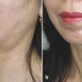
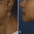
All patients get preoperative photodocumentation. We usually get an anteroposterior view while the patient is at rest, smiling, and frowning. We also get lateral and oblique views. In addition to its value in legal documentation, photography enhances communication between the surgeon and the patient.
Related posts:
 6. Facial Sculpting and Facial Slimming with Neurotoxins
 20. Comparison of Midface Rejuvenation Techniques
 22. Lower Eyelid Blepharoplasty
 5. Energy-Based Treatments for Facial Aging
 9. Male versus Female Facelift Surgery. Is There a Difference?
 12. Primary Superficial Musculoaponeurotic System (SMAS) Facelift and Neck Lift
Stay updated, free articles. Join our Telegram channel

Full access? Get Clinical Tree


