10
Atopic Dermatitis
Introduction
• Often accompanied by other atopic disorders such as asthma and allergic rhinoconjunctivitis (hay fever), which develop in an age-dependent sequence referred to as the atopic march (Fig. 10.1).

• Atopy is linked to the presence of allergen-specific serum IgE antibodies, which exist in ~70% of individuals who meet diagnostic criteria for atopic dermatitis (AD) (Table 10.1).
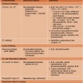
Table 10.1
Diagnostic features and triggers of atopic dermatitis (AD).


* Stroking the skin leads to a white streak that reflects excessive vasoconstriction.
URI, upper respiratory infection.
Adapted from the American Academy of Dermatology Consensus Conference on Pediatric Atopic Dermatitis (Eichenfield LF, Hanifin JM, Luger TA, et al. J. Am. Acad. Dermatol. 2004;49:1088–1095).
Clinical Features and Disease Stages of AD
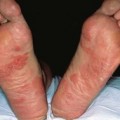
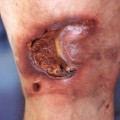
• Pruritic eczematous lesions are often excoriated and exist on a spectrum of acuity:
– Subacute lesions: erythematous patches or plaques with scaling and variable crusting.
• Regional variants of AD are depicted in Fig. 10.2.

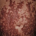
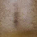
• Post-inflammatory hyper-, hypo-, or (in severe cases) depigmentation may be seen upon resolution of AD lesions (Fig. 10.3).


Fig. 10.3 Post-inflammatory pigmentary alteration in atopic dermatitis. A Ill-defined hypopigmented patches with variable residual erythema and scale on the shoulder and upper arm. B Post-inflammatory depigmentation in a patient with widespread, severe disease and multiple lesions of prurigo nodularis. A, Courtesy, Thomas Bieber, MD, and Caroline Bussman, MD; B, Courtesy, Jean L. Bolognia, MD.
• DDx: outlined in Table 10.2.

• AD is divided into infantile, childhood, and adolescent/adult stages with characteristic morphologies and distributions (see Fig. 10.2).
Infantile AD (Age < 2 Years)
• Usually develops after 6 weeks of age and often features acute lesions (Fig. 10.4).

Fig. 10.4 Atopic dermatitis on the face of an infant. Acute lesions with oozing and serous crusting are common in this age group. Courtesy, Julie V. Schaffer, MD.
• Frequently begins on the cheeks (see Fig. 10.4), forehead, and scalp; also favors the extensor aspects of the extremities and trunk (Fig. 10.5).

Stay updated, free articles. Join our Telegram channel

Full access? Get Clinical Tree


