CHAPTER 16 Vulvar Pruritus and Lichen Simplex Chronicus
The skin and modified mucous membranes of the vulva are amply endowed with slow-conducting, unmyelinated sensory nerve fibers that terminate within the upper dermis and lower epidermis. When triggered, these nerve fibers carry either pain or itching to the sensory cortex. It is not completely understood why stimulation sometimes conveys pain and, at other times, itching. However, it has recently been shown that there is a subset of these nerve fibers that are physiologically restricted to the conduction of itching. Since the most common triggers for the stimulation of these nerve endings are the environmental factors of heat, sweating, friction, and other forms of mild trauma, it is easy to see why women are more likely to experience vulvar pruritus than pruritus at most other body sites.
When women present with the symptom of vulvar pruritus, clinical examination may or may not reveal abnormalities. Occasionally, early in the course of the problem, the vulva appears entirely normal. Most often, however, there is visible evidence of scratching and rubbing. Sometimes, as indicated above, there may also be changes suggesting the presence of an underlying condition such as candidiasis or one of the vulvar dermatoses. Only the primary form of itching will be reviewed in this chapter. Secondary pruritus, associated with underlying disorders, will be discussed as these conditions are covered elsewhere in the book. Nevertheless, it is extremely important to understand that treatment for itching and scratching is the same in both the primary and secondary setting. For that reason it is permissible and appropriate to initiate antipruritic therapy even when the two types of pruritus cannot initially be separately identified.
The terms “atopic dermatitis” and “atopic eczema” were originally only used when there was clinical evidence of atopy (allergic rhinitis, hayfever, or asthma) present in the patient and/or the immediate family. Later, the definition of atopy also included elevated total immunoglobulin (Ig) E, elevated antigen-specific IgE, and eosinophilia. Because of this restricted use, in instances where the eruption had the same clinical characteristics as atopic dermatitis but where atopy could not be identified, the term “neurodermatitis” was used instead. However “neurodermatitis” carries the connotation of psychological disturbance that may or may not be present. Moreover it is a term often troubling to patients. Increasingly the atopic and the nonatopic forms of primary pruritus are now being described as “extrinsic” (i.e., allergic) atopic dermatitis and “intrinsic’ (i.e., nonallergic) atopic dermatitis1.
Lichen simplex chronicus
Epidemiology and clinical manifestations
Pruritus, especially of the anogenital region, is one of the most common conditions encountered in both men and women. In a clinic devoted to vulvar disease, itching was the single most frequent presenting symptom, occurring in 70% of patients2. In another vulvar specialty clinic, “dermatitis” was found in 25% of patients and was, after lichen sclerosus, the second most common condition encountered3. And, in an audit of 114 nonneoplastic vulvar biopsies, lichen simplex chronicus was identified in 35% of the specimens and was the most frequent condition diagnosed4.
Patient history
In any event, when itching leads to scratching, the fingernails further stimulate the cutaneous nerve endings. This increases the sensation of itching and leads to even more scratching. In short order a vicious cycle of itching and scratching develops. The presence of this “itch–scratch” cycle is highly characteristic for the presence of lichen simplex chronicus and can be viewed as its single most defining characteristic.
Psychological factors play an important part for many patients with lichen simplex chronicus5,6. Few patients volunteer the presence of anxiety and/or depression but, when directly asked, many will admit that their itching and resultant scratching are much worse when they are under stress. Based on long-term experience, I believe that these factors are frequently underrecognized and that they should be inquired about, and addressed if found, in all patients with lichen simplex chronicus.
Examination
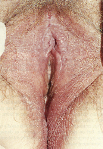
Lichen simplex chronicus, as an eczematous disease, is characterized by the standard morphology of eczematous diseases: poorly marginated, red, scaling papules and plaques (Figure 16.1). Epithelial disruption in the form of excoriations, weeping, and crusting is present initially; lichenification and pigmentary alteration develop in patients with long-standing disease.
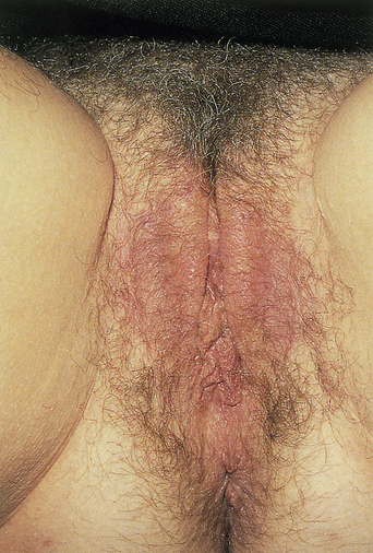
Figure 16.1 Red, lichenified, poorly demarcated plaques that are classic for lichen simplex chronicus.
(Reproduced from P. J. Lynch and L. Edwards Genital Dermatology (Fig. 5-1), New York: Churchill Livingstone; 1994.)
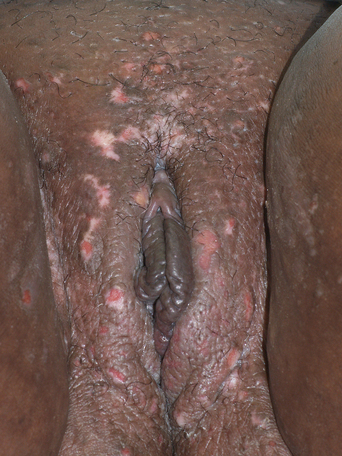
The papules and plaques are bright red initially but develop a dusky, brown-red color as time passes. Note, however, that in dark-skinned individuals, the red color may be masked by the patient’s normal brown pigmentation (Figure 16.2). The margination of the lesion, that is, the transition from abnormal to normal tissue, occurs somewhat gradually, resulting in indistinct margination. Scale is present but, because of the moisture normally present in the anogenital region, it may be visually inapparent. In this situation the scale may be only detectable as a slight roughness on palpation. As the course of the lichen simplex chronicus becomes more chronic, the surface, when moist, may whiten (see below) (Figure 16.3).
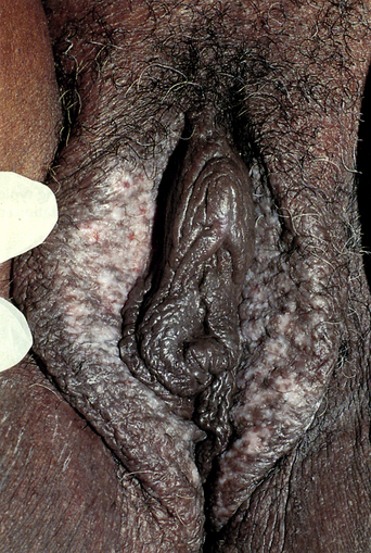
Excoriations are superimposed on the underlying inflamed red plaques and they may be superficial or deep. These scratch marks can be clinically differentiated from other causes of erosions and ulcers by their angular and linear shapes (Figure 16.4). Excoriations are accompanied by exudation (“weeping”) that leaves slightly yellow or even blood-colored stains on underwear. As the water portion of the exudate evaporates, the plasma proteins remain in place and form crusts. These crusts may be yellow or, if blood is also present, they may have red, blue, or black hues. Crusts almost always contain bacteria but a decision as to whether or not the bacteria represent infection, as opposed to colonization, must be made on other clinical evidence.