CHAPTER 25 Vulvar Edema
Edema represents the abnormal presence of fluid deep in the skin and/ or in the subcutaneous tissue. The fluid responsible for edema is either plasma (“angioedema”) or lymph (“lymphedema”). Angioedema occurs as a result of capillary dilation and excess plasma leakage into the surrounding tissue. This generally occurs as part of an acute inflammatory process and, at least in the early stages of the process, it may be transient and episodic. Lymphedema develops when there is interference with the removal of tissue fluid due to absence, destruction, or blockage of lymph flow. When angioedema due to inflammation becomes chronic, lymphatic vessel destruction occurs and there is a gradual developing blend of both angioedema and lymphedema.
Acute vulvar edema
Acute angioedema develops over minutes to hours and lasts one to several days. Resolution is accompanied by a complete return to normal-appearing skin. Many instances of acute angioedema are due to allergic reactions (medication reactions, contact allergy, etc.) but others are not (e.g., hereditary angioedema, edema due to use of angiotensin-converting enzyme inhibitors).
Allergic vulvar edema
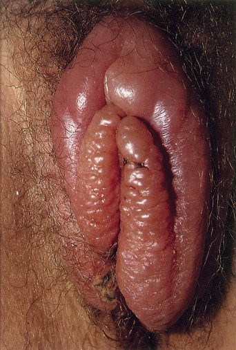
Angioedema of the vulva regularly occurs as a part of generalized allergic reactions but these patients will not present with the primary complaint of vulvar swelling. Less often, women will develop acute edema localized only to the vulva (Figure 25.1). These patients generally develop their vulvar swelling as a result of an allergic contact reaction. Two such antigens, latex and semen, are of particular importance because they are mediated by immunoglobulin (Ig) E reactions and thus may have the potential to develop anaphylaxis. Patients with reactions to either of these antigens appear to be atopic more frequently than would otherwise be expected.

Figure 25.1 Remarkable acute allergic edema which resolved spontaneously over 2 weeks.
(Reproduced from P. J. Lynch and L. Edwards Genital Dermatology (Fig. 10-14), Churchill Livingstone.)
Latex allergy occurs in about 1% of the general population and up to 4% of health care workers1. This would suggest that there is a large pool of women potentially susceptible to reactions from condoms, diaphragms, and gloved examining fingers. Exact data are lacking regarding the frequency with which vulvar problems arise secondary to latex reactions but the identification of three positive skin test reactions in a group of 92 women investigated for the presence of vulvar contact allergy suggests that this may be a commonly overlooked diagnosis2.
Approximately 80 women have been reported to develop reactions to their male partner’s semen3. But this probably underestimates the real prevalence of this condition based on the finding of two positive skin test reactions among 92 women with suspected vulvar allergic reactions2.
Many patients develop vulvar edema due to lubricants, spermicides, and medications as part of allergic contact dermatitis. These patients experience a type 4, T-cell-mediated reaction rather than a type 1, IgE-mediated reaction. Thus they develop a much greater degree of inflammation than those with classical IgE-mediated allergic reactions and generally present with an eczematous morphology. Such problems are covered in Chapter 18.
Nonallergic acute vulvar edema
Acute vulvar edema also occurs in the setting of multiple vulvovaginal infections, notably that due to candidiasis (Figure 25.2). The development of acute vulvar edema in a patient who has concomitant malaise, fever, and chills may be the earliest sign of impending necrotizing fasciitis and thus represents a medical emergency4.


