Chapter 10 Vesiculobullous disorders
1. What is the difference between a vesicle and a bulla?
If the blister is less than 5 mm in diameter, it is referred to as a vesicle; if it is 5 mm or larger, it is called a bulla. Some dermatologists require that blisters be 1 cm before using the term bulla. The term vesiculobullous disorder is often used clinically since some patients may have both vesicles and bullae.
2. How are the bullous diseases defined?
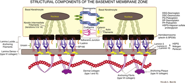
Bullous diseases are characterized by blisters. Blisters are defined as circumscribed skin lesions containing fluid. They may arise at various depths in the epidermis and dermis and are sometimes classified on the basis of the depth of skin involved. One broad classification divides blisters into those that develop within the epidermis (intraepidermal) versus those that develop below the epidermis (subepidermal) (Table 10-1). Some blistering disorders develop because of autoantibodies directed against a component of the epidermis or basement membrane zone, or they develop because of structural defects of these components. Refer to Fig. 10-1 for the location of these components in normal skin as they are discussed during questions.
4. How do you approach a patient who presents with an acute onset of a vesiculobullous eruption?
The patient history is very important in the initial evaluation of blisters. If the onset of lesions was acute, exposure to contact allergens, arthropods, phototoxic and other drugs or chemicals, trauma, and infectious agents should be queried. Certain chronic vesiculobullous diseases may have an acute onset but may then persist or recur and become chronic (Table 10-2).
5. Which skin findings are helpful in evaluating a patient with blisters?
Several features of vesiculobullous lesions are important to note, including the distribution, symmetry, involvement of mucosal surfaces, and associated lesions (such as erosions, ulcers, and crusts). Additional types of skin lesions, such as urticarial lesions, should be noted. In bullous pemphigoid, urticarial lesions often precede the development of blisters. In some vesiculobullous diseases such as dermatitis herpetiformis, secondary excoriations may be the only lesions visible, with no intact blisters.
INTRAEPIDERMAL BLISTERS | SUBEPIDERMAL BLISTERS |
---|---|
ACUTE | CHRONIC |
---|---|
The character of the blisters also may provide useful information. Flaccid blisters may indicate a more superficial blistering process than is seen with tense blisters. However, factors other than the depth of the blister are important, including site (blisters on acral skin, which has a thick stratum corneum, are often tense even when superficial) and the specific disease process (in toxic epidermal necrolysis, the blistering is subepidermal, but vesicles and bullae are usually flaccid with large sheets of skin sloughing).
7. Which tests are most useful in evaluating vesiculobullous diseases?
Most of the tests helpful in determining the cause of vesiculobullous eruptions are performed on the blister itself. When infectious causes are being considered, appropriate cultures (aerobic bacteria, viruses, fungi) may be obtained, and smears from the blisters may be stained for bacteria, dermatophytes, or the multinucleate giant cells of herpes virus infections. For noninfectious vesiculobullous diseases, a skin biopsy is often a useful test.
8. How should a skin biopsy of a vesiculobullous eruption be performed?
The lesion for biopsy should be an early lesion, to avoid secondary changes that might make the diagnosis more difficult. A small, intact blister is a good choice, as the entire lesion and some of the surrounding skin can be removed in one piece. If a punch biopsy technique is used, it is important to avoid rupturing the blister. A small excisional biopsy is a good choice and minimizes the possibility of rupturing the blister. The specimen should be placed in 10% formalin and processed for routine histologic examination. Clinical information, including the age and sex of the patient, a description of the lesions, associated symptoms, and any exacerbating factors, should be provided, along with a differential diagnosis based on the clinical examination.
9. When are special tests necessary to diagnose blistering diseases of the skin?
In addition to routine histology, a skin biopsy for direct immunofluorescence is often helpful in diagnosing the immunobullous diseases (Table 10-4). Direct immunofluorescent technique uses fluorescent, tagged antibodies that are directed against IgG, IgA, IgM, C3, and fibrin; these antibodies fluoresce when illuminated with a fluorescent microscope (Fig. 10-2). For precise diagnosis of the inherited forms of epidermolysis bullosa, electron microscopy studies may be necessary. Other tests are indicated in specific circumstances, such as urine porphyrin tests when porphyria cutanea tarda is being considered, and zinc levels when acrodermatitis enteropathica is possible.
DISEASE | CHARACTERISTIC DISTRIBUTION |
---|---|
Acrodermatitis enteropathica | Acral, periorificial |
Allergic contact dermatitis | Reflects pattern of contact; often linear |
Bullous dermatophyte infection | Feet, hands |
Bullous diabeticorum | Distal extremities |
Bullous pemphigoid | Flexural areas, lower extremities |
Cicatricial pemphigoid | Eyes, mucous membranes |
Dermatitis herpetiformis | Elbows, knees, buttocks |
Erythema multiforme | Acral areas, palms, soles, mucosa |
Hailey-Hailey disease | Intertriginous areas, neck |
Hand, foot, and mouth disease | Mouth, palms, fingers, soles |
Herpes zoster | Dermatomal distribution |
Linear IgA bullous dermatosis (childhood type) | Groin, buttocks, perineum |
Pemphigus vulgaris | Oral mucosa, other sites |
Pemphigus foliaceus | Head, neck, trunk |
DISEASE | TARGET ANTIGEN |
---|