20.2 Pitfalls
In order to enable the diagnosis of vasculitis, the choice of the “best” lesion is crucial [1–5]. A lesion of cutaneous vasculitis should be analyzed within the first 48 h after its appearance; otherwise, typical signs of vasculitis may be absent. A fresh purpuric lesion displays within the first 24 hours fibrin deposits in the vessel wall, neutrophilic infiltration, surrounding hemorrhage, and intranuclear debris.
After 24 h, lymphocytes and macrophages replace neutrophils.
After 48 h, lymphocytes predominate.
Moreover, skin biopsy of an infiltrated lesion must include the epidermis, dermis, and hypodermis to precisely determine the size of the affected vessels. Some CV affects typically the upper part of the dermis like HSP. Therefore, a punch skin biopsy will permit to show the lesions. In the case of polyarteritis nodosa (PAN), deep muscular vessels of the dermis-hypodermis and the hypodermis are affected which imply a deep incisional biopsy. Similarly, a livedo should be biopsied on its most infiltrated or necrotic areas with similar deep incisional biopsy [7].
In some specific cases, an incidental vasculitis may be found on the skin biopsy. This pathologic statement should not mislead to diagnose a vasculitis:
Biopsy performed on an ulcer
Biopsy in lesions related to neutrophilic dermatoses (Sweet’s syndrome)
20.3 Clinical Pathologic Correlation
The cutaneous lesions correlate sometimes with the size of the affected vessels [1–5]:
Palpable purpura, infiltrated erythema, urticaria, vesicles, and blisters are mainly related to small-vessel vasculitis of the dermis.
Subcutaneous nodules, ulceration, and gangrene are related frequently to medium-sized vessel vasculitis located at the dermo-hypodermal junction or in the subcutaneous fat.
Necrosis and livedo occur when either small and/or larger vessels are involved.
20.4 Clinical Manifestations
Cutaneous vasculitis displays a wide range of elementary lesions that may be associated and lead to a pleomorphic appearance of the eruption [1–5]. CV may manifest variously as:
Atypical urticaria, with distinctive feature from common urticaria (duration of the lesions longer than 24 h, presence of purpura, postinflammatory pigmentation or ecchymoses, and symptoms of burning rather than itching).
Palpable purpura: the most frequent manifestation but nonspecific; asymptomatic or burning; localized on the lower limbs, ranging from tiny red macules and pinhead- to coin-sized petechiae, but also sometimes to more extensive plaques and ecchymoses; may disclose a necrotic evolution leading to vesicles, blisters, erosions, ulcerations, and ulcer. It is often an association of different lesions simultaneously in the same patient: erythematous to purpuric macules, papules, and necrotic lesions.
Retiform purpura is a peculiar clinical form of branching purpuric lesions in a fishnet pattern for which distinction from an infiltrated or necrotic livedo is difficult. Retiform purpura implies the performance of a skin biopsy like any infiltrated purpura or livedo.
Other manifestations: infiltrated erythema; hemorrhagic vesicles; ulcers; inflammatory, tender, or painful dermal or hypodermal nodules; livedo racemosa; infarcts; and digital gangrene. Lesions affect primarily the lower limbs. Upper extremity, trunk, head, and neck involvement are not usual and may be considered as a sign of severity and/or of a systemic vasculitis (Figs. 20.2, 20.3, and 20.4).
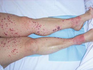
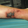
Fig. 20.2
Vasculitis of the lower limbs associating different clinical lesions of purpura: macules, papules, and vesicular lesions
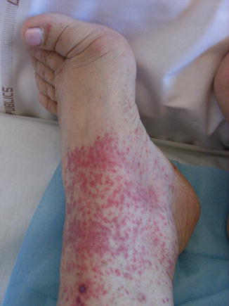
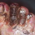
Fig. 20.3
Purpuric macules of the dorsum of the foot. Notice the absence of lesions due to the compression of the shoes
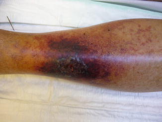
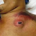
Fig. 20.4
Necrotic purpuric patch of the leg. Vasculitis was confirmed on the cutaneous biopsy
Other skin manifestations associated with systemic vasculitis but do not display vasculitis upon histology [1]:
Extravascular necrotizing granuloma: occurs during Churg-Strauss syndrome especially, red to purple papules or nodules involving symmetrically the extensor aspects of the elbows and the fingers, but other localizations have been reported.
Panniculitis: recurrent crops of erythematous, edematous, and tender subcutaneous nodules; usually of symmetrical distribution on the thighs and the lower legs; spontaneous regression with hypopigmentation and atrophic scar due to fat necrosis (lobular panniculitis) or of the extensor aspects of the lower limbs with a spontaneous regression without atrophic scar (septal panniculitis).
Pyoderma gangrenosum.
Granuloma: granulomatous lesions with neither vasculitis nor central necrosis may be observed in systemic vasculitis, especially WG with a highly variable presentation ranging from papules, nodules, subcutaneous infiltration, and pseudotumor to chronic ulcers and affecting any site of the body.
Superficial thrombophlebitis.
Gangrene resulting from arterial occlusion may be observed in all vasculitis involving medium- or large-sized arteries.
Raynaud’s phenomenon: classically associated with all types of vasculitis. However, its prevalence is unknown in many vasculitis, and its diagnostic value is very low.
20.5 Classification
The classification of vasculitis is a real brainteaser [1]. The existence of overlapping clinical features, lack of knowledge regarding the precise etiopathogenic process of each vasculitis, and lack of “pathognomonic” clinical or laboratory or radiological findings make almost impossible to have a perfect classification. Several classifications have been proposed, each of them presenting advantages and weaknesses. The most commonly used criteria for classification of vasculitis are the American College of Rheumatology (ACR) criteria established in 1990 [8] and the Chapel Hill Consensus Conference (CHCC) in 1992 [9] and revised in 2012 [10]. Conversely, the CHCC definitions – based on pathological considerations – exclude small-vessel involvement in polyarteritis nodosa (PAN). However, classification criteria should be restricted to their primary use, i.e., stratify uniform populations who carry a diagnosis. In clinical practice, a final diagnosis should rely on the interpretation of clinical, laboratory, radiologic, and pathological findings.
Related posts:


Stay updated, free articles. Join our Telegram channel

Full access? Get Clinical Tree


