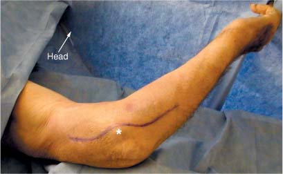
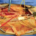
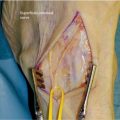
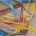
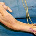
6 The ulnar nerve descends in the arm posterior to the pectoralis major muscle and medial or posteromedial to the brachial artery. At the inferior border of the pectoralis major the nerve moves medially and pierces the medial intermuscular septum ~8 cm above the medial epicondyle.1 It descends medially on the anterior surface of the medial head of the triceps muscle and then enters the interval between the medial epicondyle of the humerus and the olecranon. As it descends, the nerve is usually invested by some triceps fibers that are called the arcade of Struthers. The nerve passes into the ulnar groove on the dorsal aspect of the medial epicondyle, at the entrance to the so-called cubital tunnel. In passage through the cubital tunnel, the nerve passes from the extensor surface of the arm to the flexor surface of the forearm. As the nerve exits the tunnel, it does so between the two heads of the flexor carpi ulnaris. It lies on the palmar surface of the flexor digitorum profundus and travels in this position down through the middle of the forearm. In the distal third of the forearm it becomes superficial, lying just radial to the flexor carpi ulnaris. After emerging from under the flexor carpi ulnaris the nerve enters the wrist radial to the pisiform bone. In the forearm the ulnar nerve innervates the flexor carpi ulnaris and the flexor digitorum profundus I and II. Approximately 9 cm proximal to the wrist the ulnar nerve gives off the dorsal cutaneous sensory branch leaving the terminal motor and palmar sensory branches to traverse the wrist into the hand. The dorsal cutaneous nerve passes posteriorly, deep to the tendon of the flexor carpi ulnaris, pierces the deep fascia, and continues distally along the dorsomedial side of the wrist. It supplies sensation to the dorsal surface of the ulnar innervated fingers; that is, the fifth digit and medial half of the fourth digit. The remainder of the ulnar nerve continues into the wrist, traveling with the ulnar artery and vein as it passes lateral to the pisiform bone. The nerve fibers cross beneath the pisohamate ligament and the fibrous arch of the hypothenar muscles. It then splits off into sensory palmar digital nerves to the fifth and fourth digits, motor branches to the hypothenar muscles, deep branches to the interossei muscles, and a communicating branch to the median nerve. The ulnar nerve in the hand supplies motor function to the abductor digiti minimi, opponens digiti minimi, and flexor digiti minimi muscles of the fifth digit as well as the lumbricales III and IV. It also supplies the interossei, both dorsal and palmar, and finally the adductor pollicis and flexor pollicis brevis muscles. See Chapter 4 for the related positioning and exposure of the ulnar nerve in the arm. The patient is placed in the supine position with the arm on an arm board. The shoulder is abducted and externally rotated. The hand and forearm are fully supinated and the elbow is gently flexed (Fig. 6-1). A small ipsilateral shoulder roll may help facilitate positioning. The surgeon works from a position between the abducted arm and the patient’s body. A seated position is often the most comfortable for operating. The medial epicondyle and olecranon are marked. Exposure of the nerve within the cubital tunnel begins with a skin incision 5 cm proximal to the medial epicondyle that curves gently up and over the epicondyle. This curvature avoids placing the incision directly over the joint flexion. The incision then continues distally onto the forearm for another 4 to 5 cm. The skin and subcutaneous tissue are divided after being infiltrated with 1% lidocaine with epinephrine in a 1:100,000 concentration (Fig. 6-2). The medial antebrachial cutaneous nerve or its anterior superior branch may cross the operative field. These nerves should be identified and protected if encountered. After the skin and subcutaneous tissue are retracted, the nerve is located beneath an investing fascia within the cubital tunnel. The nerve can then be traced both proximally and distally as needed (Fig. 6-3). Distally in the cubital tunnel Osborne’s band may be encountered. It is composed of connective tissue that originates at the heads of the flexor carpi ulnaris muscle.
ULNAR NERVE
ANATOMY
In Arm and Forearm
In Wrist
POSITIONING AND SURGICAL EXPOSURE
In Arm
In Elbow and Forearm
Ulnar Nerve
Only gold members can continue reading. Log In or Register to continue

Full access? Get Clinical Tree


