Damaged venous valves result in varicose or spider vein formation. Commonly, venous obstruction is caused by increased pressure of reverse blood flow within the superficial venous valve or from direct traumatic injury to the vein.
Venous valve failure creates high pressure within the venous system which causes other valves to fail. Venous valve failure causes dilation within the entire venous system.
Venous thrombosis obstructs outflow and eventually destroys the valves within the venous system.
Introduction
Varicose veins and spider veins are normal veins that have dilated under the influence of increased venous pressure.
There are three kinds of veins in the legs- the superficial veins, which lie closest to the skin, the deep veins, which lie in groups of muscles and perforating veins, which connect the superficial veins to the deep veins. The deep veins lead to the inferior vena cava, which runs directly to the heart. In normal veins, one-way valves direct the flow of venous blood upward and inward as the leg veins must work against gravity to return blood to the heart. Varicose veins occur in the superficial veins in the legs.
One-way valves, in the veins keep blood flowing in the right direction. When the leg muscles contract, the valves inside the veins open. When the legs relax, the valves close. This prevents blood from flowing in reverse, back down the legs. The entire process is also called the venous pump.
Deep veins and perforating veins are usually able to withstand short periods of increased pressures. However, in a susceptible individual, the veins can stretch if one repeatedly sits or stands for a long time. This stretching can sometimes weaken the walls of the veins and damage the valves resulting in Varicose veins or their milder form, Spider veins.
In patients with dialysis shunts or with spontaneous arteriovenous malformations, normal veins may dilate and become tortuous in response to continued high pressure. Deep vein thrombosis initially produces an obstruction to outflow, but in most cases the thrombosed vessel eventually recanalizes and becomes a valve-less channel delivering high pressures from above downward.
Most commonly, superficial venous valve failure results from excessive dilatation of a vein from high pressure of reverse flow within the superficial venous system. Valve failure can also result from direct trauma or from thrombotic valve injury. When exposed to high pressure for a long enough periods, superficial veins dilate so much that their delicate valve leaflets are no longer able to meet.
The most common situation is that a single venous valve fails and creates a high-pressure leak between the deep and superficial systems. High pressure within the superficial system causes local dilatation leading to sequential failure (through over-stretching) of other nearby valves in the superficial veins. After a series of valves have failed, the involved veins are no longer capable of directing blood upward and inward. Without functioning valves, venous blood flows in the direction of the pressure gradient: outward and downward into an already congested leg.
Recruitment phenomena may occur, increasing the number of failing valves and communicating the high pressure into a widening network of dilated superficial veins. This may lead to a large numbers of incompetent superficial veins with the typical dilated and tortuous appearance of varicosities, over a period of time.
The deeper veins that are confined within the fascial planes remain invisible despite the fact that they can carry massive amounts of blood at high pressures. On the other hand, even a small increase in pressure can eventually produce massive dilatation of an otherwise normal superficial vein that carries very little flow. Therefore, visible varicosities are not reliable indicators of venous reflux.
The etiology of varicose veins can be classified as the following three groups:
Primary: Valvular insufficiency of the superficial veins, most commonly at the saphenofemoral junction.
Secondary
Mainly caused by deep vein thrombosis (DVT) that leads to chronic deep venous obstruction or valvular insufficiency. Long-term clinical sequelae from this have been called the postthrombotic syndrome.
Catheter-associated DVTs are also included.
Pregnancy-induced and progesterone-induced venous wall and valve weakness worsened by expanded circulating blood volume and enlarged uterus compresses the inferior vena cava and venous return from the lower extremities.
Trauma
Congenital: This includes any venous malformations. A few examples are listed as follows:
Klippel-Trenaunay variants
Avalvulia
High serum levels of estradiol are associated with clinical evidence of varicose veins in women. The hormonal changes during pregnancy may render the vein wall and valves more pliable. The sudden appearance of new dilated varicosities during pregnancy still warrants a full evaluation because of the possibility that these may be new bypass pathways related to acute deep vein thrombosis.
The relationship between serum sex steroid hormones and varicose veins in men is unclear. In a study by Kendler et al., elevated serum estradiol and testosterone levels were detected in men with varicose veins and reflux in the Great Saphenous vein (GSV) compared with the patient’s own arm veins.1 Enzymes and hormonal receptors involved in steroid metabolism were down-regulated in patients with GSV reflux and varicose veins, suggestive of a negative feedback regulation. These data support the notion of a possible causal relationship between sex steroids and varicose veins in men. The sequelae of venous insufficiency are related to the venous pressure and to the volume of venous blood that is carried in a retrograde direction through incompetent veins.
Patients may have a host of symptoms, but they are usually caused by venous hypertension rather than the varicose veins themselves. Often patients desire treatment of the unsightly nature of the tortuous, dilated varicosities only due to cosmetic reasons. Complaints of pain, soreness, burning, aching, throbbing, heavy legs, cramping, muscle fatigue, pruritus, night cramps, and “restless legs” are usually secondary to the venous hypertension. Pain and other symptoms may worsen with the menstrual cycle, with pregnancy, and in response to exogenous hormonal therapy (e.g., oral contraceptives).
On physical examination, besides, the visible palpable dilated tortuous veins, pigmentary changes may appear on the skin known as lipodermatosclerosis which results from extravasated blood. Erythematous dermatitis, which may progress to blistering, weeping, or scaling eruption of the skin of the leg or Eczema may be present and ulcers of the medial ankle most likely are the result of underlying venous insufficiency. Palpation of an area of leg pain or tenderness may reveal a firm, thickened, thrombosed vein. These palpable thrombosed vessels are superficial veins, but an associated DVT may exist in a large percentage of patients with superficial phlebitis.
The modalities available for the management of leg veins can be divided as follows:
Surgical open technique – GSV saphenectomy, Small saphenous vein (SSV) saphenectomy, Stab or ambulatory phlebectomy
Endovascular Techniques – Endovascular lasers and radiofrequency ablation
Minimally invasive techniques – Cutaneous electrodessication, Sclerotherapy
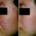
During the 1980s, lasers for leg vein treatments became available with the use of argon, though poor results were obtained due to the absence of selective photothermolysis. The development of selective photothermolysis, site-specific controlled thermal injury of tissue targets, used to treat facial telangiectasias and port-wine stains has been made available through the use of pulsed dye laser systems. Laser treatment for leg veins and telangiectasias can be selected more effectively than sclerotherapy by targeting vessels less than 1–2 mm in diameter. Ablation of the varicose pathways with the use of lasers increases the overall venous circulation. Patients with obstructed venous pathways should not undergo ablation since blood flow bypasses the obstructed areas. Longer pulse durations are used to treat large port-wine stains. Epidermal cooling devices appropriate for specified wavelength include the cryogen spray cooling system. |
Lasers for Treatment of Leg Veins
In the 1980s, leg vein treatment using lasers involved argon lasers (488 and 518 nm). The disadvantage of this laser was its strong absorption by melanin and its continuous wave nature that did not allow for selective heating of vessels, causing scarring.2,3 Continuous running Nd:YAG lasers were also tried for leg veins but poor results owing to non-specific heating of surrounding water and large depth of penetration, were observed.4,5 With the development of the principles of selective photothermolysis, the design of pulsed dye laser (PDL) came into being which effectively treated facial telangiectesias and port-wine stains.6 Longer wavelength, second generation of PDLs (595 nm) which also had longer pulse duration (1.5 ms) was launched in 1996. These PDLs had deeper penetration, yet were only effective for vessels up to 1 mm in depth and 1 mm in width.7 To overcome this, near-infrared lasers with a bandwidth of from 750 to 1,100 nm have been used to penetrate further. The longer wavelength alexandrite, diode and Nd:YAG permit sufficient energy to heat deeper leg veins up to 3 mm wide.
Indications
Many patients benefit from a combination of treatments because external lasers and light sources do not effectively treat associated reticular and varicose veins. Although sclerotherapy remains the standard treatment of leg veins and telangiectasias, lasers can be effective in treating vessels less than 1–2 mm in diameter resistant to sclerotherapy and telangiectatic matting which can occur post-sclerotherapy.
Elevated venous pressure most often is the result of venous insufficiency due to valve incompetence in the deep or superficial veins. Varicose veins are the undesirable pathways by which venous blood refluxes back into the congested extremity. Ablation of the varicose pathways invariably improves overall venous circulation.
Chronically increased venous pressure can also be caused by outflow obstruction, either from intravascular thrombosis or from extrinsic compression. In patients with outflow obstruction, varicosities must not be ablated because they are an important bypass pathway allowing blood to flow around the obstruction. Specific diagnostic tests can distinguish between patients who will benefit from ablation of dilated superficial veins and those who will be harmed by the same procedure.
An increasing popularity of lasers for treatment of leg veins has come about due to many reasons,8 especially due to its use in:
Non-cannulizable microtelangiectasias
Vessels that are refractory to conventional sclerotherapy treatments
Zones of caution such as the ankles and feet where a high incidence of complications such as hyperpigmentation and ulceration occur
Vessels that arise from prior surgical or sclerotherapy treatment (telangiectatic matting or angiogenic flushing)
Needle phobic patients
Most recently, non-surgical eradication of the greater or lesser saphenous vein (GSV and LSV, respectively).9
Contraindications
Patients with venous outflow obstruction should not have their varicosities ablated because they are important bypass pathways that allow blood to flow around the obstruction.
Technology
The laser treatment of leg veins is based on the treatment of selective photothermolysis which can be described as the production of site-specific, controlled, thermal injury of microscopic pigmented tissue targets by selectively absorbed pulses of radiation.10 It takes into account three concepts:
1.
A wavelength chosen for preferential absorption by the intended tissue chromophore.
2.
Pulse duration shorter than the thermal relaxation time (TRT). (Thermal relaxation time is defined as the time required for the target tissue to cool to half of its peak temperature after being irradiated.)
3.
Fluence high enough to cause thermal injury to the desired skin structure should be used.
As the target chromophore for laser in treating vascular lesions is oxyhemoglobin, its absorption peaks of 418, 542 and 577 nm is of importance. Also, there is a less selective peak in the range of 750–1,100 nm.11 This allows the use of PDL and IPL in treating leg veins. The longer wavelengths (700–1,100 nm) although less selective, have better penetration into the dermis, heating the entire vessel circumference and vein closure. Shorter wavelengths, on the other hand, only heat the anterior vessel wall leading to incomplete thrombosis.12 But, it is also important to keep in mind that, wavelengths greater than 900 nm could target water due to low specificity and thus a higher fluence may be needed. This could cause damage to surrounding tissues.
In order to heat leg veins, longer pulse durations than those that are used to treat port-wine stains are required as the former are comparatively larger in diameter. As most abnormal veins are 0.1–4 mm in diameter and a typical 1-mm vessel has a TRT of 360 ms, any pulse duration less than 180 ms could be used. Since a leg vein is a non-uniform target, pulse duration longer than the TRT will be needed (Altshuler’s extended theory of selective photothermolysis).13 In clinical practice, pulse durations of 10–100 ms are used. Longer pulse durations are less likely to induce vessel rupture and side effects. The early hypothesis was that by heating the blood so that the steam bubbles formed and then collapsed causing vessel rupture, also known as Cavitation. The current hypothesis believes that the vessel damage can occur either by vessel contraction secondary to collagen shrinkage or by thrombosis followed by inflammation and fibrosis.12 Heat shock protein and transforming growth factor β (TGF-β) may be mediators of collagen remodeling, fibrosis and ultimately vessel destruction. Heating of blood vessel induces formation of methemoglobin (met-Hb) in blood due to oxidative changes, and met-Hb could result from both oxygenated hemoglobin (Hb-O2) and deoxygenated hemoglobin (Hb).14–16 Once the met-Hb forms, heme protein has distorted formation and thus protein denaturation occurs. Met-Hb formation leads to change in blood absorption after laser irradiation, the maximum being at 72°C. Met-Hb has an absorbance of 4.75 times higher than that of Hb-O2 and 20 times more than that of Hb. It has been demonstrated that a series of nonuniform laser pulses with subthreshold fluences when used for closing leg veins, the first pulse induces Met-Hb formation thus leading to improved absorption by the following two pulses.17 In a study by Black et al., it was demonstrated a conformational change of the red blood cells to spheroid shape and presence of Met-Hb in blood irradiated with 1,064 nm laser at therapeutic fluences.18
Spot sizes determine how much energy reaches the desired target. Larger spot sizes cause less scatter and more energy can be available for heating the target tissue. Thus with longer wavelength, smaller spot sizes are adequate and safer. The beam diameter or spot size should be matched to the vessel diameter to maximize absorption by the vessel and minimize side effects.
Epidermal cooling techniques appropriate for each wavelength and device should always be used during laser treatment of leg veins. Various modalities can be employed such as contact cooling with a sapphire window or copper plate, cryogen spray cooling, convection air cooling, cold gels etc. These help lower the epidermal temperature while allowing the development of a peak temperature in the dermal vessels.
Lasers for Leg Vein Treatment
Potassium Titanyl Phosphate Laser (KTP Laser)
The KTP laser is a great choice for vascular lesions, especially for bright red vessels. Five hundred and thirty-two nanometer wavelength is well-absorbed by oxygenated hemoglobin and its penetration depth is not more than 0.75 mm. This appears to be great for treating superficial capillaries. But, its absorption by melanin led to frequent hyper- and hypo- pigmentation. KTP laser has showed moderate efficacy in treating veins less than 0.7 mm in width with 33% having complete response, 40% showing visible decrease in vessel diameter and 27% showing no change. Larger diameter vessels did not respond.19 The KTP laser is effective for fine-caliber vessels, with their dyspigmentation causing ability in mind. Larger spot sizes (3–5 mm) and longer pulse durations of 10–50 ms at fluences of 14–20 J/cm,2 have achieved better results.20
To use the KTP laser for leg vein treatment, a fluence of 12–20 J/cm2 and with a spot size of 3–5 mm in diameter is required, to deliver a series of pulses over the vessel until spasm or thrombosis occurs. For leg veins smaller than 1 mm in diameter that are not directly connected to a feeding reticular and with a tip chilled to 4°C to protect the epidermis, this can be an effective method. Two to three treatments may be necessary for maximal vessel improvement. Patients with darker or tanned skin may have a relatively high risk of temporary hyperpigmentation or hypopigmentation.
Long Pulsed Dye Laser
As already mentioned in brief, the 595 nm PDL was developed to overcome the limitations of the original PDL to treat the leg veins. Purpura encountered with this PDL was reduced by using its modification of a longer pulse width. Doubling the number of subpulses within each pulse led to eight consecutive subpulses that would stretch over 40 ms. On the basis of the studies performed, it is known that great results can be achieved with PDL to treat leg veins and the commonly noted side effects were edema, purpura and erythema.21 Small vessels demonstrate better results.22 Long PDL using multipass technique was tried by Tanghetti and Sherr who concluded that purpuric doses of PDL were required for the effective treatment of leg veins and hyperpigmentaion was noted in more than half of the patients treated.23
Thus, the 595 nm PDL is an effective treatment for smaller caliber (<1 mm) vessels, and the new longer pulsed systems may be even more efficacious. The major disadvantages of this laser is the inability to treat larger veins and its side effects of hyperpigmentation and purpura.
Long Pulsed 755 nm Alexandrite Laser
Long pulsed alexandrite laser in its application for leg vein treatment, provides deeper penetration into tissue and has the ability to treat larger diameter and deeply located vessels. Although this wavelength is less absorbed by hemoglobin when compared to 532 nm and 595 nm wavelengths, a sufficient level of photocoagulation of a wide range of vessel sizes can be achieved using higher fluences.
Mc Daniel et al. investigated this laser without cooling and used various parameters in 28 patients.24 According to them, the ideal fluence was 20 J/cm2 and pulse duration of 20 ms. After three sessions of 4 weekly treatments, medium vessels ranging from 0.4 to 1 mm achieved clearance of 48%. Telangiectasias less than 0.4 mm responded poorly and side effects such as bruising, erythema, crusting and hypopigmentation were noted.
In another study by Kauvar and Lou with the aim to examine the safety and efficacy of a pulsed alexandrite laser for treatment of leg telangiectasia and reticular veins, cryogen cooling was added to the protocol.25 This produced excellent clearance of telangiectasia and reticular veins of the leg with minimal adverse effects.
It can be understood that cooling devices may enable higher fluences to be used with the alexandrite laser and provide a good option for the treatment of medium vessels in the patients with lighter skin types. Severe and persistent telangiectatic matting has been reported as adverse effects.26
Diode Lasers
Related posts:


Stay updated, free articles. Join our Telegram channel

Full access? Get Clinical Tree


