(1)
Yotsuya Medical Cube, Chiyoda-ku, Tokyo, Japan
Basic Principles
Historically, areas that can be used for harvesting of vascular pedicled bone transplants include the ribs, scapula, ilium, radius, femur and fibula; however, the easiest of these to use are the fibula and the scapula. If a long bone of 7–8 cm or greater is required, then the fibula can be combined with a latissimus dorsi musculocutaneous flap or scapular flap, and in cases where only a small amount of bone but a large amount of soft tissue is required, the scapula is selected.
The PIP joint of the second toe is used for reconstructing PIP joints of the finger.
There is also a method of transferring a pedicled IDIP joint to the PIP joint, but in most cases there is a requirement that the digital arteries on both sides are patent. When harvesting from the second toe, although it is possible to retain the toe when harvesting the pedicled PIP joint, in cases where a large amount of bone needs to be harvested, there can often be problems with the remaining toe after surgery, so it is better to remove the second toe, with the consent of the patient.
Selectable Flaps and Surgical Procedures
FibulaScapulaIliumRadiusCraniumTransfer of PIP joint of second toeThe difficulty level of each surgical procedure is shown subsequent to the procedure title (e.g., Level of Difficulty: 2). The levels range from 1 to 5, with level 1 indicating a preliminary level and level 5 indicating a very advanced level.
23.1 Vascularized Fibula Graft to Lower Leg (Level of Difficulty: 5)
Information
Vascular pedicle Peroneal blood vessels
Size Can harvest around 20 cm of the fibula, leaving around 5 cm each end. Can harvest a skin flap from the middle to the distal part of the fibula with a width of 8 cm and a length of 15 cm
Anastomotic vessel Can be inserted in between the posterior tibial artery with a flow through type used with anastomosis conducted at both ends, or end-to-side anastomosis conducted with the posterior tibial artery in the popliteal region, or a side to side anastomosis conducted in the medial malleolus region.
Refer to Section of
23.1.1 Operation Procedures
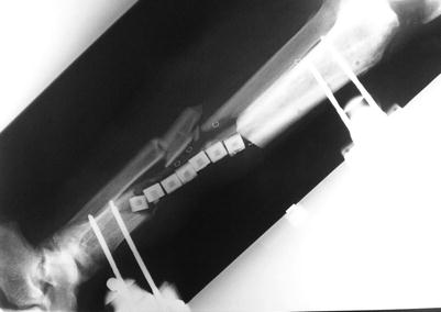
Fig. 23.1
Procedure 1: With respect to chronic osteomyelitis of the tibia following an open fracture of the lower leg, the infected bone was debrided and sustained release antibiotic hydroxyapatite inserted into the wound
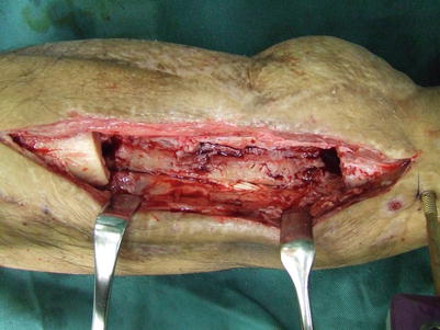
Fig. 23.2
Procedure 2: Appearance after removal of hydroxyapatite. An 8 cm bone defect is present
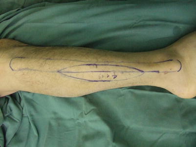
Fig. 23.3
Procedure 3: The fibula flap is designed in the center of the lower leg
Refer to Section of
“Bone and joint transplant/Vascular pedicle free fibular transplant to the forearm” in Chap. 22
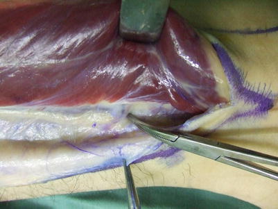
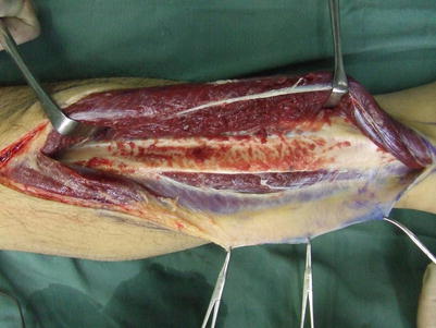
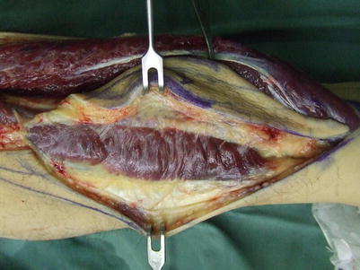
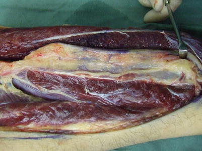
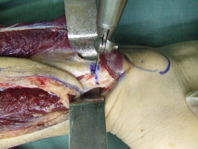
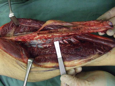
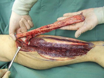
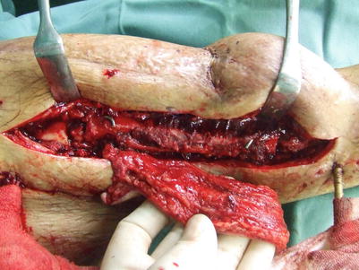
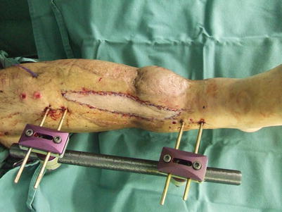
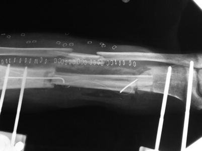
Fig. 23.4
Procedure 4: An incision is made in the anterior margin of the fibula flap, and the perforators to the flap are confirmed as emerging from behind the bone. The peroneal muscle is pulled forward and dissected
Fig. 23.5
Procedure 5: The anterior fibula is detached
Fig. 23.6
Procedure 6: The posterior margin of the flap is opened and with part of the soleus fascia in the flap, it is detached from the muscle
Fig. 23.7
Procedure 7: While confirming and retaining the perforators, detachment is continued until the flexor hallucis longus muscle comes into view
Fig. 23.8
Procedure 8: A retractor is inserted into the posterior side of the distal fibula, and the bone is severed while protecting the vascular pedicle
Fig. 23.9
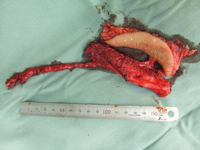
Procedure 9: The vascular pedicle running along the posterior surface of the bone is ligated, and while holding onto the bone, the vessel running along the muscle is detached, with about 1 cm from inside the flexor hallucis longus muscle included. The proximal end of the bone is cut, and further dissection conducted
Fig. 23.10
Procedure 10: The posterior tibial muscle is cut from the anterior tibia. The peroneal blood vessels are detached proximally up to the branch with the posterial tibial blood vessels, however care is taken not to damage the sural nerve behind the head of the fibula
Fig. 23.11
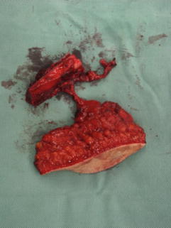
(a, b) Procedure 11: The harvested pedicled fibula and flap. The bone is folded in two while retaining the vascular pedicle
Fig. 23.12
Procedure 12: The tip of the bone is shaved, inserted into the marrow at both ends and fixed in place with a steel wire. Side-to-end anastomosis is conducted of the vascular pedicle to the posterior tibial blood vessels
Fig. 23.13
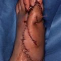
Procedure 13: A continuous suction tube is inserted under the flap and the flap is sutured
Fig. 23.14
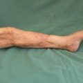
Procedure 14: Appearance of transplanted bone fixed in place
Tips
Which to choose, free vascularized bone graft or bone transport method?
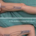
A fibula that cannot be folded in half is too thin and prone to fracture after commencement of load bearing. The transplanted fibula can become thicker due to the fracture, but it will take another several months of treatment before it can take a full load. In the bone transport method that uses the Ilizarov external bone fixation and extension apparatus, ossification and leg extension can be conducted simultaneously following resection of a nonunion section. Although it takes time, it is a stable process, and capable of handling thick bones and soft tissue defects.
Related posts:
Stay updated, free articles. Join our Telegram channel

Full access? Get Clinical Tree


