Tibiotalocalcaneal and Pan-Talar Arthrodesis

CHAPTER 38 Tibiotalocalcaneal and Pan-Talar Arthrodesis


OVERVIEW


Whenever possible, it is preferable to perform a tibiotalocalcaneal (TTC) arthrodesis instead of a pan-talar arthrodesis. Including the transverse tarsal joint in the arthrodesis, which results in far more rigidity to the foot, is rarely necessary. When performing a talectomy and a tibiocalcaneal (TC) arthrodesis, I leave the navicular bone free of the anterior aspect of the remnant of the talus. With some deformities, the entire talus must be removed, and the anterior tibia will then abut the navicular. In patients with neuropathy and a Charcot deformity, including the navicular bone in this arthrodesis is tempting, because it will increase the surface area for the arthrodesis. In this manner, after a cheilectomy of the anterior distal tibia and debridement of the navicular, a tibionavicular arthrodesis can be added to the TC arthrodesis. In general, however, I do not consider this additional fusion to be advisable, because the function of the foot, particularly in the setting of a neuropathy, is better without it. Thus the only indications for a pan-talar arthrodesis are to address severe pan-talar arthritis and to correct a deformity of such magnitude that a TTC fusion alone will be inadequate.


The surgical approach for exposure in a pan-talar arthrodesis is almost identical to that for a combined ankle arthrodesis–triple arthrodesis. In fact, I start the procedure with exposure of the ankle joint using the mini-arthrotomy approach with two anterior incisions and then extend these distally once the ankle is completely debrided. The medial incision is extended distally from the ankle to expose the talonavicular joint. Laterally, however, if the incision for the ankle arthrodesis is extended distally, then one incision is placed slightly anterior or dorsal to the calcaneocuboid joint. A fibulectomy should not be performed, if possible. The same rationale applies with a pan-talar arthrodesis as with the ankle arthrodesis: Preservation of the fibula is desirable because the blood supply to the ankle is maintained. Likewise, preservation of the medial malleolus is preferable, although at times, correction of ankle deformity cannot be accomplished without either an osteotomy or a resection of the medial malleolus. An important case in point is that of a TTC arthrodesis with intramedullary (IM) rod fixation, in which slight medial translation of the foot under the tibia is helpful. This translation can be accomplished only if the medial malleolus is removed.


For fixation of the pan-talar arthrodesis, the approach is similar to that outlined for the screw technique used for a TTC arthrodesis. The only difference is that here the fixation of the talonavicular joint can be extended proximally into the tibia with screws started at the inferior pole of the navicular bone, which cross both the talonavicular and the tibiotalar joints into the back of the tibia.


TIBIOCALCANEAL AND TIBIOTALOCALCANEAL ARTHRODESIS


Fixation Alternatives


The fixation options for a TC arthrodesis are screws, a blade plate, and an IM rod. To some extent, the choice of fixation may depend on personal preference, but at times a more stable fixation with either a blade plate or an IM rod is preferable in the setting of significant bone loss and deformity. In my experience, when any erosive changes or avascular necrosis of the ankle is present, screws may not be strong enough, particularly with neuropathic deformity. Historically, a blade plate has been demonstrated to be biomechanically superior to an IM rod in torsion and bending strengths, but this finding was reported with use of an inferior IM rod device; by comparison, the current rod designs include capability for internal and external compression as well as locking mechanisms for the distal screws to create a fixed-angle device similar to the blade plate.


Thus, with the newer designs of the IM nail, rod fixation is preferable to use of a blade plate, provided that sufficient calcaneal bone is present. If the calcaneal bone is of poor quality, the posterior-to-anterior screw can be inserted across the calcaneocuboid joint into the cuboid bone. Postoperative weight-bearing status also may be a consideration. In patients in whom compliance with a non–weight-bearing regimen is in question, I prefer to use an IM rod that can be dynamized if necessary. Although the locking screws may break, a nonunion is not as worrisome in the setting of neuropathic deformity, provided that the foot remains axially aligned under the tibia.


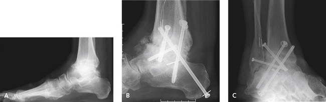
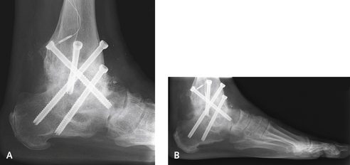
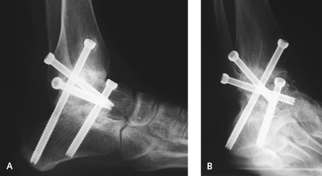
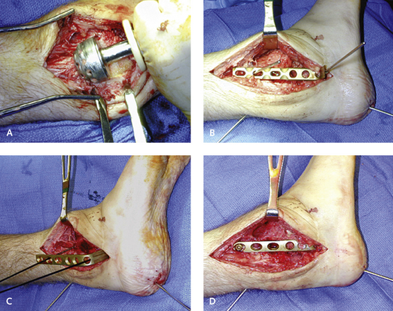
I do sometimes use screws alone for a TTC fusion, but only when the bone quality is good, the alignment of the limb is not significantly abnormal, and focal arthritis is present without bone loss or avascular necrosis (Figures 38-1 to 38-4). I rarely use screws alone when the talus is missing and the overall alignment of the limb is not satisfactory. At present, therefore, for a TTC arthrodesis, I use screws for correction of minimal deformity in ankles with good bone quality and an IM rod in a majority of the remaining cases. A blade plate remains useful when tibia deformity is present, if an IM rod cannot be used or if a simultaneous tibia osteotomy cannot be performed for realignment.


Surgical Approaches


A lateral transfibular approach to the ankle and hindfoot can be used for correction of severe deformity. Although this distal fibulectomy will devascularize the lateral ankle, an alternative often is unavailable for correction of severe deformity, particularly when the ankle is angulated and in varus and the fibula is prominent. The incision is made vertically, directly over the fibula, extending down distally over the sinus tarsi toward the inferior aspect of the calcaneus. The sural nerve must be identified and then retracted inferiorly with the peroneal tendons. A fibulectomy is performed with an acetabular reamer, which is used to completely denude and decorticate the fibula. The reamings that are obtained are preserved for later use as bone graft material (Figure 38-5).


If a blade plate is to be used for fixation, the distal 8 cm of the fibula must be removed, and following use of the reamer, the fibula is cut with a saw. I prefer to use a chisel and not a saw to denude the ankle and subtalar joints. If severe deformity is present, however, then the distal tibia may have to be cut at the plafond with a saw. Often the foot cannot be centered under the ankle because the medial malleolus blocks the shift of the talus. In such instances, the malleolus is removed with an oblique osteotomy made through a separate medial incision. The dissection in the sinus tarsi and subtalar joint is performed in the same manner as that described for a subtalar arthrodesis.


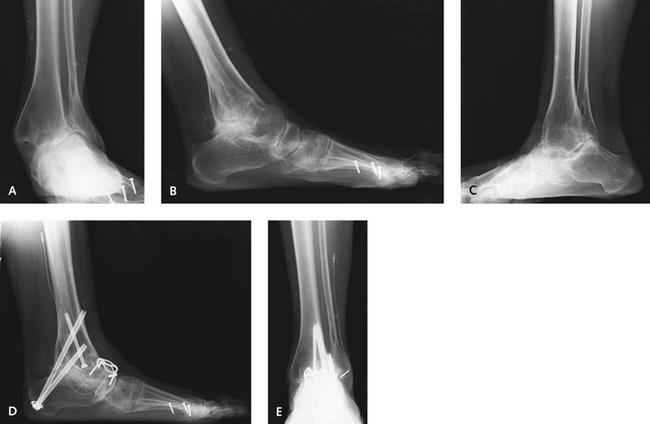
When a talectomy plus a TC arthrodesis is performed for avascular necrosis, the necrotic remnants of the talus are completely excised. A sizable defect remains; either it can be filled with bone graft or the talus can be apposed directly onto the calcaneus (Figure 38-6). Usually, despite the contouring of the posterior aspect of the calcaneus and the undersurface of the distal tibia, joint apposition cannot be easily obtained. It is difficult to appose the calcaneus flush up against the tibia, because the hindfoot tilts up into dorsiflexion, leaving the hindfoot in a calcaneus position. A defect of variable size, depending on bone erosion, is always present between the undersurface of the tibia and the dorsal surface of the posterior facet; the shape of the defect may range from a trapezoid to a large triangle (Figure 38-7). At times, if the apposition between the tibia and the calcaneus is very good and the defect is not too large, it can be filled with cancellous graft only. Some bone graft is needed in the more anterior aspect of the arthrodesis to fill the defect properly. Here either cancellous graft or a tricortical structural allograft can be used; the choice will depend on the availability of large structural graft and the size of the defect. It is clearly easier to secure the posterior tibia to the dorsal calcaneus and then fill the defect with cancellous graft. Over the past several years, with the increased use of orthobiologic adjuvants, the latter has been my preferred technique (Figure 38-8). I use a structural graft when the height of the limb must be restored. In some circumstances, this structural graft has to be used between the tibia and the calcaneus. Of note, in performing a talectomy and TC arthrodesis, as the heel is pushed up against the tibia, the skin on either side of the ankle gets compressed, and closure can no longer be achieved without tension on the skin (the so-called accordion effect—as the structure is elongated, it narrows, and as it is compressed, it widens).

Only gold members can continue reading. Log In or Register to continue

Stay updated, free articles. Join our Telegram channel

Mar 6, 2016 | Posted by in Reconstructive surgery | Comments Off on Tibiotalocalcaneal and Pan-Talar Arthrodesis

Full access? Get Clinical Tree

Get Clinical Tree app for offline access