(1)
Department of Neurosurgery Faculty of Medicine, Saga University, Saga, Japan
Keywords
Cerebellar arteriesSCAAICAPICAOverviewSegmentation3.1 Introduction
The three cerebellar arteries comprise superior cerebellar artery (SCA), anterior inferior cerebellar artery (AICA), and posterior inferior cerebellar artery (PICA). SCA, AICA, and PICA course around the brainstem, near the three cerebellar peduncles, and in the three cerebellar–brainstem fissures between the cerebellum and brainstem to reach the three cerebellar hemispheres, as described in Chap. 1: “The “Rules of Three” in the Posterior Cranial Fossa” (Fig. 3.1). SCA courses around the midbrain, supplying the anterior part of the roof of the fourth ventricle, the superior cerebellar peduncle in the cerebellomesencephalic fissure, and the tentorial (superior) cerebellar surface [1, 8]. AICA courses around the pons, supplying the lateral part of the fourth ventricle, the middle cerebellar peduncle in the cerebellopontine fissure, and the petrosal (lateral) cerebellar surface [4, 8]. PICA courses around the medulla oblongata, passing through the lower cranial nerves (CNs), and supplies the inferior cerebellar peduncle in the cerebellomedullary fissure, the posterior part of the roof of the fourth ventricle, and the suboccipital (posterior) cerebellar surface [3, 8].
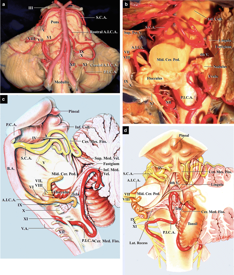
Fig. 3.1
Relationship between the three cerebellar arteries and neural structures in the posterior cranial fossa. (a) The brainstem and cerebellar arteries. Anterior view. (b) The three cerebellar arteries in the cerebellar–brainstem fissures. Left superolateral view; the left cerebellar hemisphere has been removed, and the fourth ventricle opened. SCA courses in the cerebellomesencephalic fissure, whereas PICA courses in the cerebellomedullary fissure. Consequently, SCA and PICA run on the upper and lower surfaces of the fourth ventricle, respectively. (c) Color illustration showing the relationship between the three cerebellar arteries and the brainstem and fourth ventricle. Lateral view; the left half of the cerebellum has been removed (from Matsushima T et al. [8] with permission). (d) Color illustration showing the relationship between the three cerebellar arteries and the cerebellar–brainstem fissures and fourth ventricle. Superior view; the left and part of the right halves of the cerebellum have been removed (from Matsushima T et al. [8] with permission)
In this chapter, we explain the definitions, courses, and segments of the three cerebellar arteries and their perforators. We also examine the relationship between these arteries and CNs. Precise anatomical knowledge of the cerebellar arteries is essential for planning a preoperative surgical strategy and for safe, successful surgery in the posterior cranial fossa.
3.2 The Superior Cerebellar Artery
3.2.1 Definition
There are two possible definitions of SCA. One defines SCA as the artery with the most distal origin among the infratentorial cerebellar arteries. Another defines SCA as the artery that supplies the tentorial cerebellar surface. Because the former definition is more specific, it has been adopted as the definition of SCA.
3.2.2 Overview
SCA originates from the basilar artery (BA) near its apex. It dips caudally and encircles the brainstem near the pontomesencephalic sulcus. After passing CN V, SCA enters the cerebellomesencephalic fissure, where its branches supply the superior cerebellar peduncle and are finally distributed to the tentorial cerebellar surface (Fig. 3.2a–e). SCA usually arises as a single trunk, but may occasionally arise as a double trunk in 15–30 % of cases.
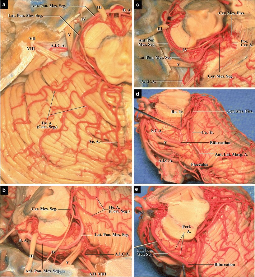
Fig. 3.2
The superior cerebellar artery, cerebellomesencephalic fissure, and tentorial cerebellar surface. (a) Overview of the superior cerebellar artery (SCA). Superior view. Left SCA passes around the midbrain to enter the cerebellomesencephalic fissure, finally reaching the tentorial cerebellar surface. (b) SCA around the midbrain. Left anterolateral view. (c) SCA and its perforating arteries in the cerebellomesencephalic fissure. Left lateral view. (d) The bifurcation of SCA and its relationship to cranial nerve (CN) V. Left lateral view. (e) The circumflex perforating arteries of SCA. Left lateral view
3.2.3 Segments
Hardy DG et al. [1] divided SCA into four segments: (1) Anterior pontomesencephalic, (2) Lateral pontomesencephalic, (3) Cerebellomesencephalic, and (4) Cortical (Table 3.1).
Table 3.1
Segments of the superior cerebellar artery
Anterior pontomesencephalic segment |
Lateral pontomesencephalic segment |
Cerebellomesencephalic segment |
Cortical segment |
The anterior pontomesencephalic (pontine) segment begins at the origin of SCA, extending laterally below CNs III to the anterolateral margin of the brainstem (Fig. 3.2a, b).
The lateral pontomesencephalic (ambient) segment begins at the anterolateral margin of the brainstem and travels along the lateral part of the pons, parallel to CN IV (Fig. 3.2c, d). SCA often bifurcates into rostral and caudal trunks in this segment (Fig. 3.2d). The rostral trunk supplies the cerebellar vermis and the medial part of the cerebellar hemisphere, whereas the caudal trunk supplies the lateral part of the cerebellar hemisphere.
The cerebellomesencephalic (quadrigeminal) segment courses within the cerebellomesencephalic fissure (Fig. 3.2c). Bilateral SCAs become proximate in this segment. This segment gives rise to the precerebellar artery, which supplies the superior cerebellar peduncle.
The cortical segment gives rise to branches distal to the cerebellomesencephalic fissure, which pass below the tentorial edge and are distributed to the tentorial cerebellar surface. Branches of the cortical segment are divided into the vermian and hemispheric groups (Fig. 3.2a). The vermian branches ascend the culmen after dipping caudally at the level of the declive and anastomose with the vermian branches of PICA on the tentorial cerebellar surface. The marginal artery is the most proximal of the cortical branches. It usually arises from the pontomesencephalic segment and courses along the anterolateral margin of the cerebellum; therefore, it is also called the anterolateral marginal artery (Fig. 3.2d).
3.2.4 Relationships to Cranial Nerves
SCA passes near CNs III, IV, and V and frequently makes contact with them (Fig. 3.2a–c). Because the proximal part of SCA passes below CN III, the nerve is sandwiched by SCA inferiorly and the posterior cerebral artery (PCA) superiorly. CN IV arises below the inferior colliculus and traverses forward in the cerebellomesencephalic fissure. On reaching the lateral side of the brainstem, it courses below the lower surface of the tentorium, penetrating the dura mater at the posterior edge of the oculomotor trigone to enter the cavernous sinus. CN IV passes superomedial to the lateral pontomesencephalic segment of SCA as it traverses forward. CN V arising from the lateral part of the upper pons exists in the upper cerebellopontine angle. It traverses forward, beneath the tentorial attachment to enter the Meckel’s cave. The lateral pontomesencephalic segment of SCA encircles the brainstem and makes contact with CN V from the superomedial side while forming a caudal loop near or on the nerve (Fig. 3.2d). Compression of CN V by SCA may cause trigeminal neuralgia. SCA has a few potential points of contact with CN V because it bifurcates into rostral and caudal trunks in addition to forming a caudally projected loop (Fig. 3.2d, e) [2, 5].
3.2.5 Perforators
The perforating branches of SCA are divided into direct and circumflex types [1]. The direct type pursues a straight course to enter the brainstem in the interpeduncular fossa. The circumflex type winds around the brainstem and supplies the tegmentum, cerebellar peduncle, and inferior colliculus. SCA gives rise to the precerebellar arteries in the cerebellomesencephalic fissure, which course along the superior cerebellar peduncle in the fissure and supply the inferior colliculus, superior medullary velum, and dentate nucleus. The superior colliculus is mainly supplied by PCA.
3.2.6 Imaging
The four segments of SCA—a downward loop formation of the anterior and lateral pontomesencephalic segments and the rostral and caudal trunks—can be visualized using magnetic resonance imaging (MRI), magnetic resonance angiography (MRA), and conventional angiography. Before the advent of MRI and MRA, it was considered that the shape of SCA in the anterior–posterior view using angiography shows that of the brainstem, and the downward loop formation of the lateral pontomesencephalic segment of SCA may be related to the contact between the artery and CN V. Nowadays, the relationships between SCA and CNs can clearly be observed using MRI and MRA, including preconstructed MRA images. In cases of trigeminal neuralgia, careful attention should be paid to the relationship between CN V and SCA because SCA is often involved in the disease.
(Regarding imaging of SCA, please refer to Chap. 10: “Anatomy for Microvascular Decompression Procedures: Relationships between Cranial Nerves and Vessels, Preoperative Images, and Anatomy for the Stitched Sling Retraction Technique”)
3.3 The Anterior Inferior Cerebellar Artery
3.3.1 Definition
Related posts:
 The Veins of the Posterior Cranial Fossa: Nomenclature
 The Cerebellopontine Angle: Basic Structures and the “Rules of Three”
 The Temporal Bone: Basic Anatomy and Approaches to Internal Auditory Canal
 Microvascular Decompression for Glossopharyngeal Neuralgia: Surgical Approaches Depending on the Offending Artery
 Microsurgical Anatomy of and Surgical Approaches to the Jugular Foramen
 Microsurgical Anatomy of the Cerebellomedullary Fissure and Variations of the Transcerebellomedullary Fissure Approach
Stay updated, free articles. Join our Telegram channel

Full access? Get Clinical Tree


