(1)
Department of Neurosurgery Faculty of Medicine, Saga University, Saga, Japan
Keywords
Veins of the posterior cranial fossaNomenclatureVeins of the tentorial (superior) cerebellar surfaceVeins of the petrosal (lateral) cerebellar surfaceVeins of the suboccipital (posterior) cerebellar surfaceVeins of the brainstem4.1 Introduction
From a radiologist’s viewpoint, Huang YP et al. divided the veins of the posterior cranial fossa into three draining groups based on the direction of drainage: the galenic, tentorial, and petrosal draining groups (Fig. 4.1) [1–4]. Although their works are excellent, their nomenclature is unsuitable for neurosurgeons. Furthermore, recognizing and remembering the names of veins in the posterior cranial fossa using their nomenclature is difficult; knowledge of the veins of the posterior cranial fossa and how they relate to systematic neural structures, surgeries, and surgical approaches is crucial for neurosurgeons who encounter them during surgery. In this chapter, we explain our classification and nomenclature of the veins of the posterior cranial fossa based on the neural structures and surgical approaches [9].
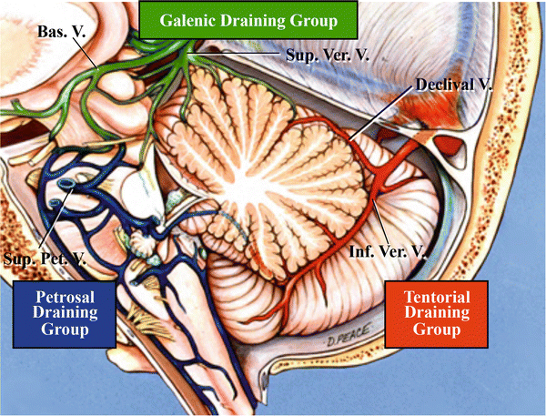
Fig. 4.1
The three draining groups of the veins in the posterior cranial fossa. Green denotes the galenic draining group; dark blue denotes the petrosal draining group; and brown denotes the tentorial draining group (from Matsushima T et al. [9] with permission)
4.2 Neural Structures and Nomenclature of Veins of the Posterior Cranial Fossa
Most major cerebellar veins course on the surfaces of neural structures. As shown on angiography by Huang YP et al., the veins of the posterior cranial fossa represent the shape of the brainstem and cerebellar hemispheres [1–4]. Description of the veins of the posterior cranial fossa requires detailed knowledge of the surface anatomy of the brainstem and cerebellum. Neural structures in the posterior cranial fossa comprise the cerebellum and brainstem, including the midbrain, pons, and medulla oblongata. Three major fissures exist between the cerebellum and brainstem [8, 9].
(Regarding neural structures in the posterior cranial fossa, refer to Chap. 2: “Neural Structures: the Brainstem, Cerebellum, Cerebellar Peduncles, and Fourth Ventricle”)
Veins in the posterior cranial fossa are divided into four groups: (1) superficial veins, (2) deep veins, (3) veins of the brainstem, and (4) bridging veins [5–9]. Superficial veins course on the cortical surfaces of the cerebellum. Deep veins run in three deep cerebellum–brainstem fissures. Veins of the brainstem course on the surface of the brainstem. Superficial veins, deep veins, and veins of the brainstem are described in this chapter, but bridging veins are described in detail in Chap. 5: “The Bridging Veins in the Posterior Cranial Fossa” because bridging veins are more relevant to surgery.
4.3 Surgical Approaches and Veins of the Posterior Cranial Fossa
We previously described a simplified nomenclature applicable to surgical approaches to the posterior cranial fossa [8, 9]. Our nomenclature is predominantly based on the relationship of these veins to the three cortical surfaces of the cerebellum and the three deep cerebellum–brainstem fissures. The three cortical surfaces of the cerebellum comprise the tentorial (superior), petrosal (lateral), and suboccipital (posterior) cerebellar surfaces. The three deep fissures, situated in the deep area of each surface, comprise the cerebellomesencephalic, cerebellopontine, and cerebellomedullary fissures. We will explain the veins of each surface, including both superficial and deep veins.
4.4 Veins of the Tentorial Cerebellar Surface
The veins of the tentorial cerebellar surface comprise superficial veins coursing on the cortical surface and deep veins coursing in the cerebellomesencephalic fissure (Table 4.1, Fig. 4.2). In addition, bridging veins draining into the Group 2 of the tentorial sinuses are also grouped with the superficial veins. We classified the tentorial sinuses on the basis of the origins and draining direction into four groups; the tentorial sinuses which drain into the torcula or the transverse sinus near it are named Group 2 [10].
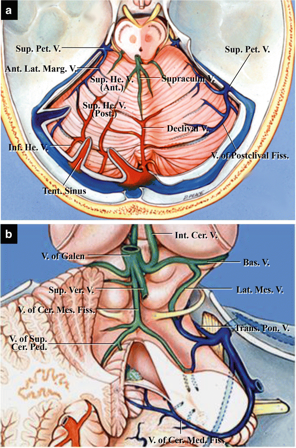
Table 4.1
Veins of the tentorial cerebellar surface
Superficial veins |
Midline―Superior vermian vein (Supraculminate vein, Declival vein) |
Lateral―Superior hemispheric veins |
Anterior group |
Posterior group |
Deep veins |
Midline―Veins of the superior cerebellar peduncle |
Vein of the cerebellomesencephalic fissure |
Lateral―Lateral mesencephalic veins |
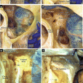
Fig. 4.2
Veins of the tentorial cerebellar surface. (a) Superficial veins; superior view. They include the superior vermian veins and the superior and inferior hemispheric veins. The superior vermian vein occasionally divides into the supraculminate and declival veins (from Matsushima T et al. [9] with permission). (b) Deep veins. They include the vein of the cerebellomesencephalic fissure and the veins of the superior cerebellar peduncle (from Matsushima T et al. [9] with permission)
The superficial veins of the tentorial cerebellar surface are divided into two groups: longitudinal and transverse veins. The latter are superficially invisible because these veins often course in the cerebellar fissures. The vein of the postclival fissure is a major transverse vein. Longitudinal veins on the surface include the superior vermian vein near the midline and the superior hemispheric veins located on the hemispheric part of the tentorial cerebellar surface. The superior vermian vein courses superiorly on the culmen and joins the vein of the cerebellomesencephalic fissure to drain into the vein of Galen. The superior vermian vein is sometimes divided into the supraculminate vein, which ascends toward the vein of Galen, and the declival vein, which descends toward the torcula. The superior hemispheric veins are divided into anterior and posterior groups. The anterior group of the superior hemispheric veins ascends toward the apex of the culmen to join the superior vermian vein. The posterior group of the superior hemispheric veins drains the posteromedial part of the tentorial cerebellar surface. The posterior group usually comprises longitudinal veins, which join the inferior hemispheric veins from the suboccipital cerebellar surface, forming the bridging veins that drain into the tentorial sinuses near the postclival fissure. The courses of the posterior group of the superior hemispheric veins and inferior hemispheric veins influence the formation of Group 2 of the tentorial sinuses.
Related posts:
 The Cerebellopontine Angle: Basic Structures and the “Rules of Three”
 Three Cerebellar Arteries: Superior Cerebellar Artery, Anterior Inferior Cerebellar Artery, and Posterior Inferior Cerebellar Artery
 The Temporal Bone: Basic Anatomy and Approaches to Internal Auditory Canal
 Microvascular Decompression for Glossopharyngeal Neuralgia: Surgical Approaches Depending on the Offending Artery
 Microsurgical Anatomy of and Surgical Approaches to the Jugular Foramen
 Microsurgical Anatomy of the Cerebellomedullary Fissure and Variations of the Transcerebellomedullary Fissure Approach
Stay updated, free articles. Join our Telegram channel

Full access? Get Clinical Tree


