(1)
Department of Neurosurgery Faculty of Medicine, Saga University, Saga, Japan
Keywords
Temporal boneFacial nerveInternal auditory canalCochleaSemicircular canalsVestibular schwannoma surgery18.1 Introduction
Surgeries through the temporal bone are challenging because of its complicating anatomy inside the bone [15–17, 21, 22]. In this chapter, to understand the basic anatomy of the temporal bone, we will describe the following:
The basic structures of the temporal bone, both for surgery and radiological interpretation
The microsurgical anatomy of the three approaches to the internal auditory canal (IAC) through the temporal bone
The important structures that neurosurgeons must have a detailed knowledge of include IAC, cochlea, three semicircular canals, and cranial nerve (CN) VII. In this chapter, we describe the relationships between these structures. (Regarding the transpetrosal approaches and surgeries related to IAC, refer to Chap. 14: “Microsurgical Anatomy of the Internal Auditory Canal and Surrounding Structures, and Vestibular Schwannoma Surgery” and Chap. 19: “Posterior and Anterior Transpetrosal Approaches.”) The approaches through the temporal bone have many variations depending on their purpose. However, they share one aim: to avoid injury of CN VII and postoperative hearing disturbance.
18.2 Basic Structures
18.2.1 Constituents: The Squamous, Petrous, and Tympanic Parts
The temporal bone contains the external ear, middle ear (tympanic membrane, tympanic cavity, three ossicles, Eustachian tube, and antrum), internal ear (vestibule, semicircular canals, and cochlea), and facial canal. The facial, cochlear, and superior and inferior vestibular nerves course inside the bone, especially within IAC (Fig. 18.1a).
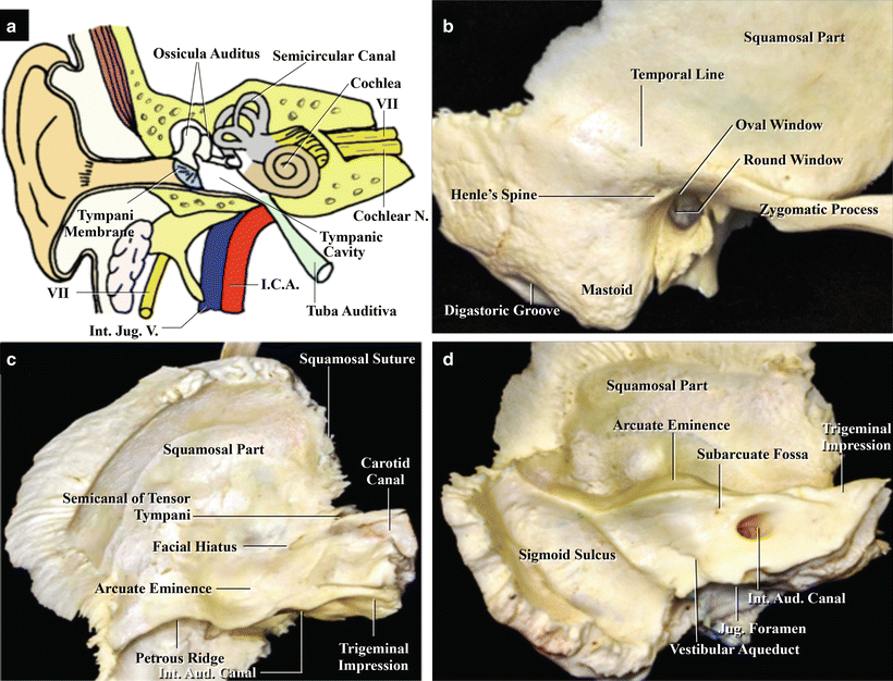
Fig. 18.1
Basic structure of the ear and the three surfaces of the temporal bone. (a) Illustration showing the structures of the right ear. Anterior view (from Matsushima T [14] with permission). (b) Dry temporal bone: lateral surface. (c) Dry temporal bone: superior surface (middle cranial fossa floor). (d) Dry temporal bone: medial surface (posterior cranial fossa lateral wall)
The temporal bone, which is joined anteriorly to the sphenoid bone, superiorly to the parietal bone, and posteriorly to the occipital bone, is comprised by three parts: squamous, petrous, and tympanic. The junction of these parts on the lateral side forms the external auditory canal (Fig. 18.1b) [21]. The squamous part—a round, bony plate standing erect sagittally—has a convex external surface, and its zygomatic process projects forward from the anterosuperior part of the porus acusticus externus to join the zygomatic bone, forming the zygomatic arch. The petrous part contains the mastoid process, which projects downward, and the petrous bone, which projects anteromedially and wedges between the sphenoid and occipital bones (Fig. 18.1c, d). The digastric groove below the mastoid process is the site of attachment of the sternocleidomastoid muscle and can be palpated behind the lower part of the auricle (Fig. 18.1b). The occipital artery and occipital nerve pass superomedially through this groove. The inner surface of the mastoid process also has a groove, called the sigmoid sulcus, in which the sigmoid sinus courses (Fig. 18.1d). The interior of the mastoid process in adults is filled with mastoid air cells, which communicate with the mastoid antrum. Therefore, the mastoid bone ultimately communicates with the external environment.
The petrous bone contains IAC, which opens as the oval porus acusticus internus (PAI) in the center of the posterior surface of the petrous bone (Fig. 18.1d). The cochlea lies anterolateral to IAC, the semicircular canals lie lateral to it, the carotid canal courses anteroinferior to it, and the jugular foramen lies posteroinferior to it.
18.2.2 Interior Structures of the Temporal Bone, with Special Reference to the Internal Auditory Canal and Surrounding Structures
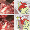
IAC is surrounded by the cochlea and three semicircular canals. After coursing in IAC, CN VII runs in the facial canal between the cochlea and semicircular canals from the posterior to middle cranial fossa, where it forms the geniculate ganglion. The nerve then runs downward in the mastoid bone toward the stylomastoid foramen. The dry bone shown in Fig. 18.2a, b demonstrates the cochlea (highlighted in red) anterolateral to IAC and the semicircular canals (highlighted in green) posterolateral to it. CN VII, highlighted in yellow, which enters the temporal bone through PAI, is also visible coursing around the mastoid antrum after passing the middle cranial fossa.
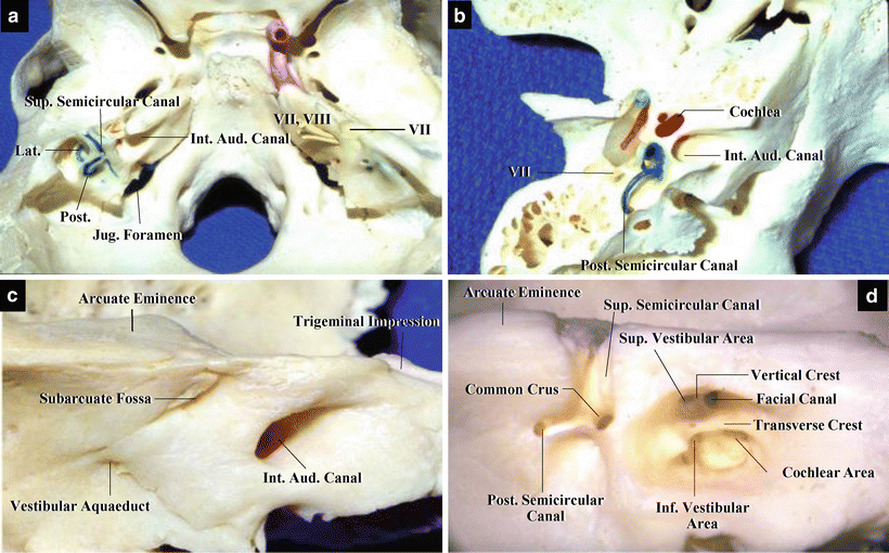
Fig. 18.2
Dry bones showing the internal auditory canal (IAC) and surrounding structures, such as the cochlea, three semicircular canals, and facial canal (from Matsushima T [14] with permission). (a) Dry skull. Superior view. The three semicircular canals have been exposed on the left side, and the facial canal has been exposed on the right side. (b) Dry left temporal bone. Superior view. IAC and surrounding structures are visible. (c) Dry left temporal bone. Posterior view. The vestibular aqueduct and subarcuate fossa are visible superolateral and inferolateral to the porus acusticus internus, respectively. (d) Dry left temporal bone. Posterior view. IAC and semicircular canals have been opened. The fundus of IAC and common crus are visible. The common crus is lateral to the fundus. The lateral end of the fundus is separated into four parts by the transverse and vertical crests
Surrounding PAI on the surface of the posterior cranial fossa are several structures relevant to neurosurgeons, including the subarcuate fossa, which contains the subarcuate artery, and the vestibular aqueduct, which communicates with the aquaeductus cochleae (Fig. 18.2c). In the dry bone shown in Fig. 18.2d, the bone covering IAC and semicircular canals have been removed, exposing the interior of IAC, superior semicircular canal, and posterior semicircular canal. The junction between the superior and posterior semicircular canals is termed the common crus. The lateral end of IAC is divided into four parts. The transverse crest divides it into the upper and lower parts, and the vertical crest further subdivides the upper portion into the anterior and posterior parts. The anterior part is the facial canal, in which CN VII passes, and the posterior part is the superior vestibular part, through which the superior vestibular nerve passes. The area below the transverse crest is also divided approximately into two areas, and the cochlear nerve and inferior vestibular nerve traverse them, respectively.
18.2.3 Imaging Anatomy
Osseous structures in the temporal bone are visible with computed tomography (CT). Axial-view CT scans shown in Fig. 18.3a, b demonstrate the structures present at the levels of the external auditory canal and IAC. Air cells, including the mastoid air cells, occupy most of the temporal bone. Malleus and incus are visible in the middle ear, and the cochlea, which is connected with them, is visible in the internal ear (Fig. 18.3a). The compact bone, which does not contain any air cells, lies posterolateral to IAC. It can be seen as a white, high-density mass on CT (Fig. 18.3b). The semicircular canals are situated inside this compact bone (Fig. 18.3b).
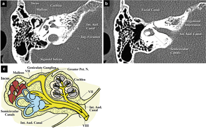
Fig. 18.3
Computed tomography (CT) imaging of the temporal bone (from Matsushima T [14] with permission). CT reveals the internal auditory canal (IAC), facial canal, cochlea, three semicircular canals, incus, malleus, and jugular foramen. (a) Left side. Superior view at the level of the ossicula auditus. (b) Left side. Superior view at the level of the facial canal from the posterior to middle cranial fossa. (c) Illustration showing the structures surrounding the left IAC. Superior view. Cranial nerves (CNs) VII and VIII run in the canal. CN VII forms the geniculate ganglion with the greater petrosal nerve on the middle cranial fossa. The cochlea is anterolateral to the canal, and the three semicircular canals are posterolateral to it
Regarding the relationships between the external, middle, and internal ears, the external auditory canal and middle ear are bounded by the tympanic membrane, and a small foramina called the round window and the oval window exist in the boundary between the middle and internal ears. The base of the stapes, an ear ossicle, fits into the oval window, and it propagates vibration from the tympanic membrane to this window (Fig. 18.3c). Vibration transmitted from the middle ear excites the hair cells of the organ of Corti (or spiral organ) in the cochlea, which transmits the stimulus to the cochlear nerve. The semicircular canals monitor equilibrioception: the semicircular canals, which contain endolymph liquid, are located at right angles to one another, thus permitting differentiation of rotational movement in all directions. The nerves in the semicircular canals and saccule combine, forming the superior and inferior vestibular nerves, and enter the medulla oblongata after merging with the cochlear nerve within IAC. The cochlear nerve from the cochlea runs in the anteroinferior portion of IAC, and the superior and inferior vestibular nerves run in the posterior half of IAC (Fig. 18.3c).
18.2.4 The Course of Cranial Nerve VII and Its Branches
Figure 18.4 shows the dry temporal bones after mastoidectomy or unroofing of IAC. CN VII is visible running from the posterior to the middle cranial fossa, then descending toward the stylomastoid foramen inside the petrous bone, running around the external auditory canal (Fig. 18.4). CN VII, which originates from the lower pons, first runs in the anterosuperior part of IAC, coursing in the facial canal toward the middle cranial fossa to form the geniculate ganglion (Fig. 18.4a, b). The parasympathetic branch, originating from the medulla oblongata and coursing together with the nerve bundle of CN VII, divides from the geniculate ganglion and joins the greater petrosal nerve. The nerve courses in the great petrosal sulcus to the facial hiatus (Fig. 18.4b). Gustatory nerve fibers, which originate from nerve cells in the geniculate ganglion, combine to form the nervus intermedius. Its central branch runs in IAC with CN VII to enter the pons. While CN VII descends in the mastoid bone, the peripheral branch divides from CN VII, joins the parasympathetic secreting fibers to form the chorda tympani, and innervates the tongue.
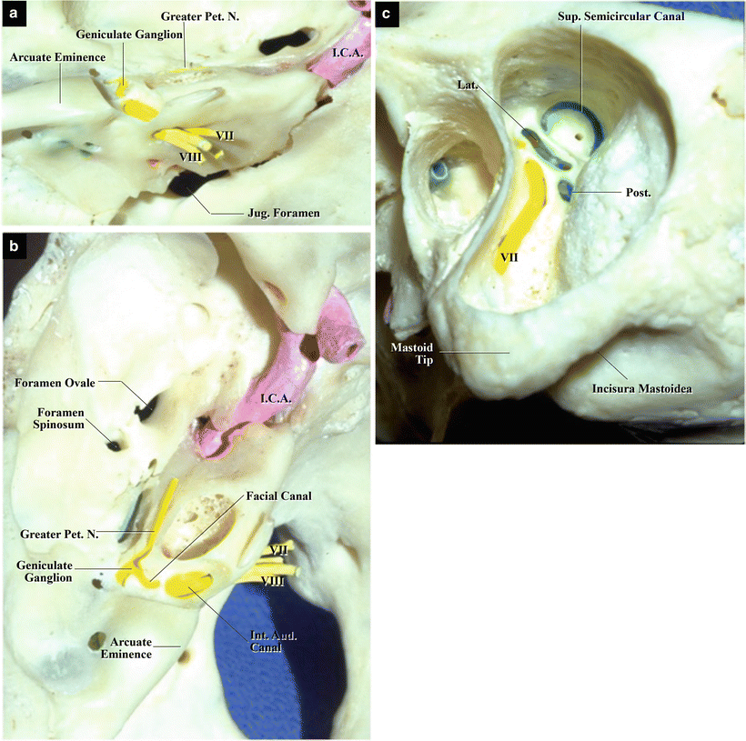
Fig. 18.4
The course of cranial nerve (CN) VII (from Matsushima T [14] with permission). The facial canal in the dry temporal bone has been opened on the left side. (a) Temporal bone showing the posterior and middle cranial fossae. Posterosuperior view. CN VII courses from the posterior to middle cranial fossa between the cochlea and semicircular canals. (b) Temporal bone showing the middle cranial fossa. Superior view. CN VII forms the geniculate ganglion with the greater petrosal nerve anterior to the arcuate eminence. (c) Temporal bone showing the interior of the mastoid process. Lateral view. Mastoidectomy has exposed the facial canal and CN VII. The nerve runs downward from the middle cranial fossa to the stylomastoid foramen around the external auditory canal
Related posts:
 The Veins of the Posterior Cranial Fossa: Nomenclature
 The Cerebellopontine Angle: Basic Structures and the “Rules of Three”
 Microvascular Decompression Surgery for Hemifacial Spasm: The Lateral Suboccipital Infrafloccular Approach
 Microvascular Decompression for Glossopharyngeal Neuralgia: Surgical Approaches Depending on the Offending Artery
 Microsurgical Anatomy of and Surgical Approaches to the Jugular Foramen
 Microsurgical Anatomy of the Cerebellomedullary Fissure and Variations of the Transcerebellomedullary Fissure Approach
Stay updated, free articles. Join our Telegram channel

Full access? Get Clinical Tree


