(1)
Department of Neurosurgery Faculty of Medicine, Saga University, Saga, Japan
Keywords
Three brainstem componentsThree cerebellar pedunclesThree cerebellar surfacesThree cerebellar-brainstem fissuresThree cerebellar arteriesThree draining groups of the veins of the posterior fossa1.1 Introduction
Each structure within the posterior cranial fossa, including the brainstem, cerebellar surfaces, cerebellar peduncles, and cerebellar arteries and veins comprises of three parts (Table 1.1). Rhoton AL Jr and colleagues proposed the “rules of three” to understand the anatomy of the posterior cranial fossa and cerebellopontine angle (CPA) [1, 4, 8, 9]. Particularly, the etiology of neurovascular compression syndromes is easily understood when related to the three anatomical components of the CPA [2, 3, 6, 7]. In this chapter the “Rules of Three” is explained.
Table 1.1
The “Rules of Three” in the posterior cranial fossa
Brainstem | Midbrain |
Pons | |
Medulla oblongata | |
Cerebellar surfaces and related surgical approaches | Tentorial (superior) cerebellar surface |
Infratentorial supracerebellar approach | |
Petrosal (lateral) cerebellar surface | |
Lateral suboccipital approach | |
Suboccipital (posterior) cerebellar surface | |
Midline suboccipital approach | |
Cerebellar peduncles | Superior cerebellar peduncle |
Middle cerebellar peduncle | |
Inferior cerebellar peduncle | |
Cerebellar-brainstem fissures | Cerebellomesencephalic fissure |
Cerebellopontine fissure | |
Cerebellomedullary fissure | |
Cerebellar arteries | Superior cerebellar artery |
Anterior inferior cerebellar artery | |
Posterior inferior cerebellar artery | |
Veins of the posterior cranial fossa | Galenic group |
Petrosal group | |
Tentorial group |
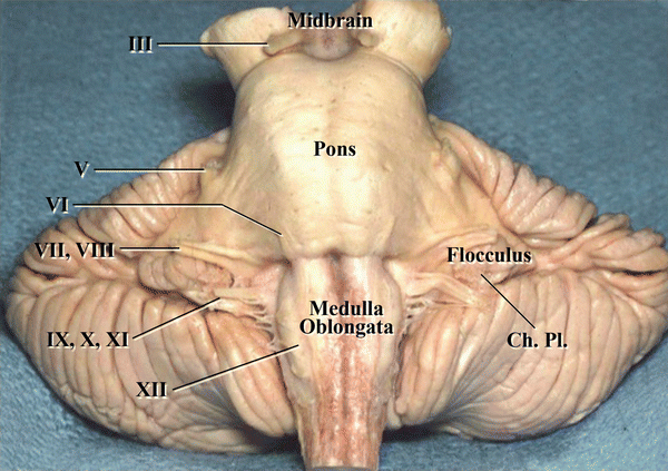
1.2 Brainstem
1.3 Cerebellar Surfaces
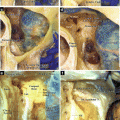
Based on the surgical approaches, the cerebellar surfaces are divided into three types as follows (Figs. 1.2 and 1.3) [6, 8, 9]:
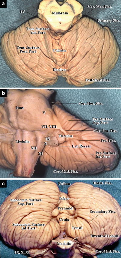
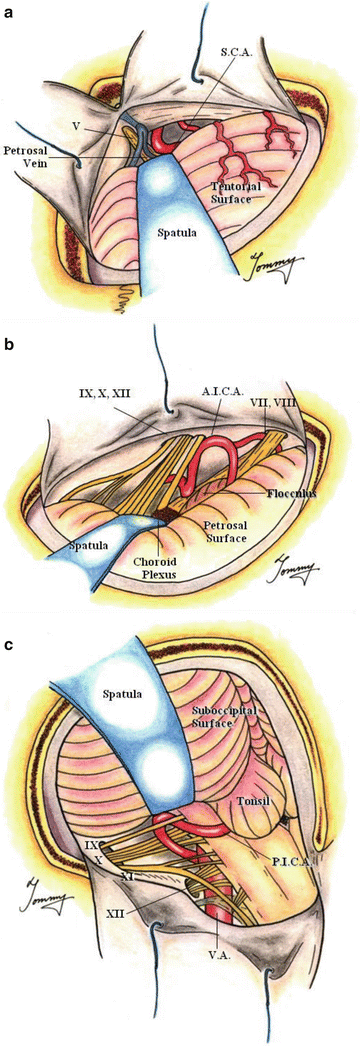
Fig. 1.2
Three cerebellar surfaces. (a) Tentorial surface, (b) Petrosal surface, (c) Suboccipital surface
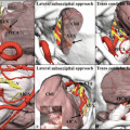
Fig. 1.3
Three cerebellar surfaces and the surgical approach to each surface (from Hitotsumatsu T et al. [3] with permission) (a) Infratentorial approach and tentorial surface, (b) Lateral suboccipital approach and petrosal surface, (c) Suboccipital approach and suboccipital surface
Tentorial (superior) cerebellar surface:
1.
 The Veins of the Posterior Cranial Fossa: Nomenclature
 The Cerebellopontine Angle: Basic Structures and the “Rules of Three”
 The Temporal Bone: Basic Anatomy and Approaches to Internal Auditory Canal
 Microvascular Decompression for Glossopharyngeal Neuralgia: Surgical Approaches Depending on the Offending Artery
 Microsurgical Anatomy of and Surgical Approaches to the Jugular Foramen
 Microsurgical Anatomy of the Cerebellomedullary Fissure and Variations of the Transcerebellomedullary Fissure Approach


Opposing the cerebellar tentorium, it is exposed via the transoccipital transtentorial or supracerebellar approach (Figs. 1.2a and 1.3a).
Related posts:
The Veins of the Posterior Cranial Fossa: Nomenclature
The Cerebellopontine Angle: Basic Structures and the “Rules of Three”
The Temporal Bone: Basic Anatomy and Approaches to Internal Auditory Canal
Microvascular Decompression for Glossopharyngeal Neuralgia: Surgical Approaches Depending on the Offending Artery
Microsurgical Anatomy of and Surgical Approaches to the Jugular Foramen
Microsurgical Anatomy of the Cerebellomedullary Fissure and Variations of the Transcerebellomedullary Fissure Approach
Stay updated, free articles. Join our Telegram channel
Full access? Get Clinical Tree
