The Rheumatoid Foot and Ankle

CHAPTER 39 The Rheumatoid Foot and Ankle


FOREFOOT RECONSTRUCTION


The standard technique for correction of rheumatoid forefoot deformity has involved resection of the metatarsal heads with realignment of the lesser toe deformities and arthrodesis of the hallux metatarsophalangeal (MP) joint. Over the years, in my experience, this has been the most reliable procedure for correction of deformity, particularly that associated with erosive changes of the MP joint with destruction of bone, especially in the metatarsal head.


Modifications of the procedure may include resection of the lesser metatarsal heads with resection arthroplasty of the hallux MP joint (as an alternative to arthrodesis). This resection-based approach is certainly an option, although it does not appear to give as stable a result as that achieved with arthrodesis. Nevertheless, the resection arthroplasty can be considered in the presence of arthritis of both the MP and the interphalangeal (IP) joints of the hallux. If the arthritis is associated with deformity of both joints, then one alternative is to perform an arthrodesis of the IP joint in conjunction with a resection arthroplasty of the MP joint. I am, however, opposed to performing a resection arthroplasty of the hallux in the patient with rheumatoid arthritis and hallux valgus, because soft tissue laxity associated with erosive changes and ligamentous instability will lead to a recurrence of deformity. Some patients with these gross deformities are not symptomatic, and their function is acceptable. Which is preferred—form or function? Certainly, if function follows form, then the resection arthroplasty of the hallux (the Keller procedure) is not an ideal procedure, because the hallux is weak and unstable and the incidence of recurrent deformity is high.


Other options for correction of the deformity at the MP joint of the lesser toes include synovectomy and shortening osteotomies of the metatarsal head or shaft. Synovectomy is certainly an option to consider in the presence of severe synovitis associated with minimal joint deformity and the absence of metatarsalgia. Once subluxation or dislocation of the MP joints occurs, however, synovectomy is not of any benefit.


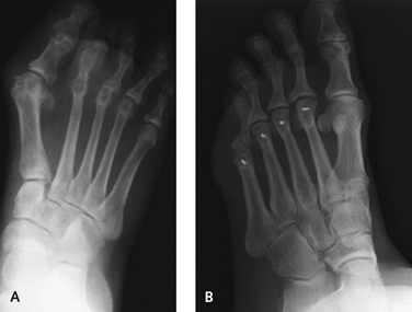
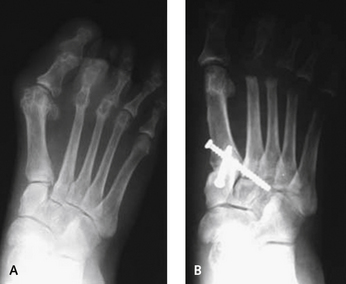
The concept of joint preservation of the MP joint is an important one in patients with inflammatory joint disease. The synovitis surprisingly decreases with mechanical off-loading of the MP joint as a result of the shortening osteotomy. Although the lesser metataral osteotomy is a reasonable procedure to be performed in a patient with rheumatoid arthritis, it is not easily accomplished because of the paucity of good bone around the metatarsal head, the erosive changes typically associated with joint subluxation and dislocation, and the difficulty of performing this operation while maintaining a salvageable joint. Nonetheless, in patients who have involvement of one or two joints without severe erosion, shortening osteotomy is an option. This procedure is indicated especially when one or two of the lesser MP joints are involved, and resection of all of the metatarsal heads is inadvisable for some reason (Figure 39-1).


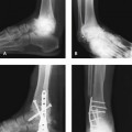
Many intermediate stages of deformity of the rheumatoid forefoot exist in which arthrodesis of the MP joint may not be considered necessary. A good example is the presence of hallux valgus in an otherwise healthy joint. In this instance, a standard operation for correction of hallux valgus (e.g., bunionectomy and metatarsal osteotomy) may not be as successful as, for example, a tarsometatarsal arthrodesis (the modified Lapidus procedure) (Figure 39-2). If hallux valgus is not present initially and metatarsal head resections are performed, then the hallux deformity will always increase as a result of shortening of the lesser toes in the absence of a lateral buttress to the hallux. Preservation of the hallux MP joint is even more relevant if joint preservation osteotomy procedures of the lesser metatarsal heads are performed.


INCISIONS AND DISSECTION


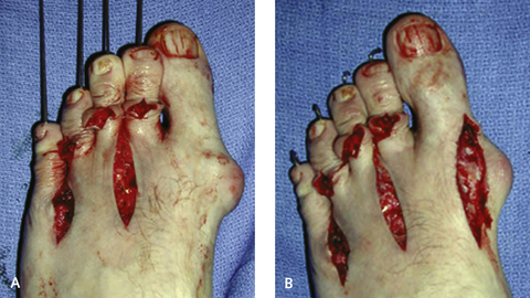
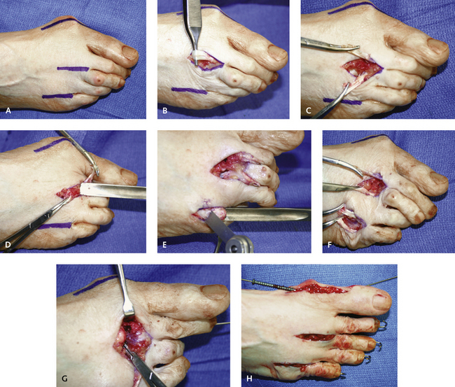
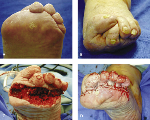
The choice of incisions used for the metatarsal head resections or the metatarsal head osteotomies is determined by the magnitude of deformity. In general, I prefer two dorsal longitudinal incisions made in the second and fourth web spaces (Figure 39-3). The improved access with these incisions, however, must be balanced against the possibility of wound dehiscence and associated problems with skin healing in this region. The option of a dorsal transverse incision is available, but not if dislocation of the MP joints is present with shortening and contracture of the soft tissues. Although the transverse incision is easier to perform and can be done with far less retraction than with two longitudinal incisions, the surgeon must be certain that sufficient bone has been resected to facilitate soft tissue closure. The other option is a plantar surface–based elliptical incision. Although I have used this incision on occasion, it is associated with problems with management of the contracted dorsal soft tissues, including the extensor tendons and capsule. I see no advantage to use of a plantar surface–based incision other than the proximity of the metatarsal heads. The hypertrophied soft tissue, callus, or bursae are always resorbed once the metatarsal heads have been resected, and the excision of an ellipse of tissue on the plantar surface does not seem warranted (Figures 39-4 and 39-5).


As noted, I generally use two dorsal longitudinal incisions made in the second and the fourth web spaces, with as wide a skin bridge as possible between them. The metatarsals are resected first, followed by the MP fusion. With this sequence, inadvertent manipulation of the hallux MP joint is avoided. Both incisions should be as long as possible and extend from the cleft of the web space proximally for approximately 4 cm. Care must be taken not to overretract the tissue, to prevent bruising and ecchymosis of the tissue during the dissection. When one side of the skin is retracted, the other is relaxed. I find it easier to start with the more lateral fourth web space incision, because the fifth metatarsal is always the easiest to remove. In each case, the extensor tendons of the toes are transected 2 cm proximal to the metatarsal neck and then clamped and pulled distally. This maneuver facilitates the exposure of the MP joint through retraction of the tendon all the way up to the dislocated joint.


The dissection is deepened, and a capsulectomy is performed until the MP joint is identified. This joint is not always easy to identify if it is dislocated, but this must be reduced before the osteotomy of the neck is performed. I use a curved periosteal elevator inserted over the dorsal surface of the MP joint; then, with plantar surface–directed pressure, the remnant of the metatarsal head is delivered into the dorsal surface of the wound. The proximal phalanx is depressed underneath the metatarsal head for further exposure. A problem encountered in this setting is the presence of soft bone, which causes crushing of the metatarsal head when retracted with the elevator. The soft bone does not create a problem with the resection of the head, but remnants of the metatarsal head must be sought after it is removed. This problem also may arise at the base of the proximal phalanx, which can be fractured; retrieval of the bone fragments may be difficult here as well. I do not resect the base of the proximal phalanx, even with severe dislocation. This resection unnecessarily shortens the toe and adds to dorsal instability.


The metatarsal neck is cut with a saw at the level of the flare between the metaphysis and the diaphysis of the metatarsal. Use of an osteotome will fracture the metatarsal in an irregular manner, creating irregular bone spikes, and is therefore not recommended. The cut is made obliquely in a slightly plantar direction, to avoid creating any plantar spike, which will lead to metatarsalgia. I use a clamp or towel clip to hold, rotate, and then pull out the head, while cutting the collateral ligaments, and remnant of the plantar plate.


What should be done with the extensor tendons? As a matter of expedience, these can be simply left cut or repaired. Neither of these choices is my preference, however, because my aim here is to prevent the development of any dorsiflexion contracture postoperatively. I therefore perform a plantar tenodesis of the extensor tendons. Each extensor tendon is grasped with a hemostat and then passed underneath the metatarsal neck. A Kirschner wire (K-wire)is then passed in antegrade and then retrograde fashion across the MP joint and through the tendons into the metatarsal itself. This wire holds the extensor tendons under the metatarsal neck, and the tenodesis effect prevents dorsal extension contracture. The K-wires are never stable enough in osteopenic bone. Stability can be maximized, however, with insertion of the K-wire as proximal as possible into the cuneiforms or cuboid bone. Wound closure is performed with nylon sutures in the skin only, because the subcutaneous tissue is rarely thick enough to hold a suture. No tension at all should exist on the skin incisions.


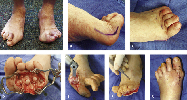
The correction of the claw toe deformities often has been described as unnecessary. Of note, however, if the toes are left deformed at the proximal IP joint, MP joint deformity tends to recur later as well. These contractures should be addressed with either manual manipulation of the joint or resection arthroplasty. Although a formal arthroplasty is my preferred procedure (Figure 39-6), manual manipulation of the joint occasionally will be successful in other than rigid contractures.


I prefer to leave the K-wires in for 6 weeks to gain as much stability as possible at the posterior interphalangeal (PIP) and MP joints. The K-wires are inserted as far posteriorly as possible, even into the cuneiforms, to prevent early loosening.


Mar 6, 2016 | Posted by in Reconstructive surgery | Comments Off on The Rheumatoid Foot and Ankle

Full access? Get Clinical Tree

Get Clinical Tree app for offline access