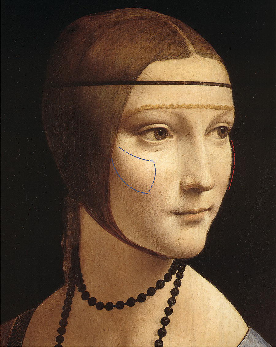
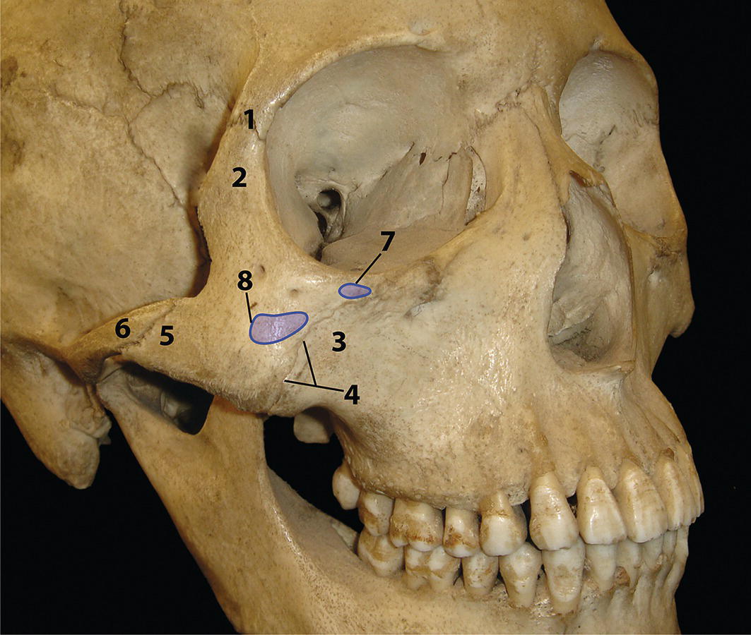
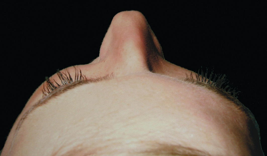
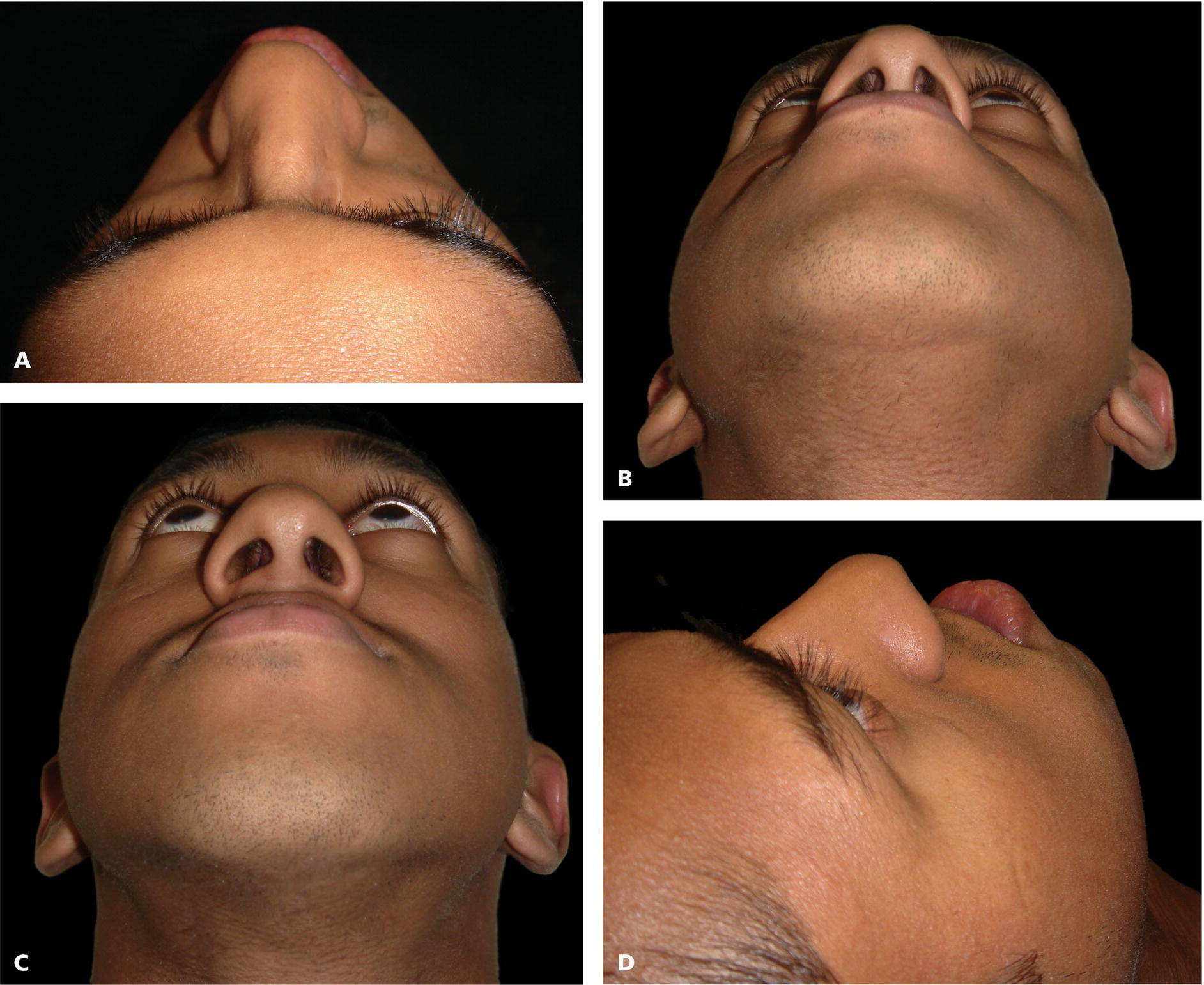
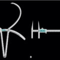
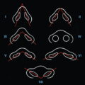
One of the most recognizable features of the midface is the contour and prominence of the superior‐lateral part of the cheek, termed the malar eminence or ‘cheekbone’, formed by the underlying zygomatic bone. The malar eminence is a recognized characteristic of facial attractiveness (Figure 15.1). Malar hypoplasia or deficiency is due to lack of projection of the body of the zygomatic bone. A number of terms may be used to describe the layperson’s term ‘cheekbone’. The terms malar bone, zygomatic bone, zygoma and os jugale are often used interchangeably, although there is only one term for the zygomatic arch. The term ‘zygomatic’ is derived from the Greek and Latin words for a ‘yoke’, which is a wooden crosspiece fastened over the necks of two animals and attached to a plough or cart that they are forced to pull. Moreover, a pair of animals may be thus ‘yoked’ together. The derivation is presumably due to the joining together of various skeletal structures by the zygomatic bone, and the modest resemblance of the bizygomatic region in frontal view to a yoke. Figure 15.1 The malar eminence is an important aesthetic feature of the face, forming the contour and prominence of the superior‐lateral part of the cheek. (Modified, detail, Lady with an Ermine, portrait of Cecilia Gallerani, Leonardo da Vinci, c. 1485, Czartoryski Museum, Kraków.) Figure 15.2 Anatomy of the zygomatic bone: The anterior and slightly convex lateral surface of the zygomatic bone forms the bony prominence of the upper cheek, also termed the malar eminence; in layperson’s terms, this is the cheekbone (Figure 15.2). It is perforated by the zygomaticofacial foramen. Two important muscles of facial expression, particularly important for smiling, arise from the zygomatic bone. Zygomaticus major arises from the anterolateral surface of the zygomatic bone, and zygomaticus minor from just superior to the zygomaticomaxillary suture. Together with a number of smaller muscles, these muscles cause a superior and lateral movement of the midfacial soft tissues associated with smiling and the expression of joy. The zygomatic arch is formed by the zygomatic process of the temporal bone and the temporal process of the zygomatic bone. Anteriorly, the superiorly projecting frontal process of the zygomatic bone and the zygomatic process of the maxilla may be considered as the anterior pillars of the arch. The zygomatic process of the maxilla is palpable through the soft tissues of the cheek and intraorally in the buccal sulcus. Figure 15.3 Right‐sided malar deficiency in a patient with hemifacial microsomia. The zygomatic region must be clinically evaluated for: The relationship of the malar regions to the rest of the patient’s face must be evaluated in frontal, oblique lateral and left/right profile views. It is useful to analyse the malar region from above and behind (superior view), and from below (inferior view). In addition, it is particularly useful to evaluate malar projection, or lack thereof, from an oblique superior profile view (Figure 15.4). Photographic records may be taken from all these views. The bizygomatic width should be the widest region of the face in frontal view. The bizygomatic width and its relation to the facial height, bitemporal width and bigonial width, has a significant influence on overall facial form (Table 15.1). The facial height to width ratio (facial index) gives the overall facial type, such as ‘tall’ or ‘short’ or ‘square’ face. The proportionate facial height (nasion‐menton) to bizygomatic width ratio is approximately 90% for men and 85% for women. Figure 15.4 Evaluation of malar region: (A) superior view; (B) inferior view; (C) oblique inferior view; (D) superior oblique lateral view. Table 15.1 Facial widths and total face height in white Caucasians Normative values based on original data from Farkas1.
Chapter 15
The Malar Region
Introduction
Terminology

Anatomy
Clinical evaluation
Bizygomatic width
Male (mm)
Range (mm)
Female (mm)
Range (mm)
Bitemporal width (Ft’‐Ft’)
115
111–121
110
108–116
Bizygomatic width (Zy’‐Zy’)
140
134–144
130
125–135
Bigonial width (Go’‐Go’)
105
99–113
95
90–100
Face height (N’‐Me’)
125
119–130
110
107–116
Total face height (Tr‐Me’)
190
175–199
170
165–181
Related posts:


Stay updated, free articles. Join our Telegram channel

Full access? Get Clinical Tree


