(1)
Department of Neurosurgery Faculty of Medicine, Saga University, Saga, Japan
Keywords
Cerebellopontine angleCranial nervesCerebellar arteriesPetrosal veinsInternal auditory canal8.1 Introduction
Detailed knowledge of the surgical anatomy of the cerebellopontine angle (CPA) is vital for surgical treatment of CPA tumors and vertebral artery (VA) aneurysms and for microvascular decompression (MVD) surgeries [4, 7, 8, 11, 13, 18–20, 23]. CPA is a cistern that is surrounded by the petrous bone laterally and the petrosal (lateral) cerebellar surface and brainstem medially. Many cranial nerves (CNs), arteries, and veins run in this cistern [17, 22]. First, the lateral and medial walls of CPA will be described; later, CNs and vessels, especially the basic relationships between nerves and arteries, will be explained. The microsurgical anatomies of the internal acoustic meatus and lateral part of the foramen magnum are described in detail in Chaps. 14 and 17, respectively. The microsurgical anatomy and surgeries for MVD are described in Chaps. 10–13.
8.2 The Lateral Wall of the Cerebellopontine Angle
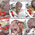
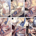
The lateral wall of CPA is formed by the temporal and occipital bones. Their borders, the petro-occipital fissure, form the inferior petrosal sulcus, which is a groove housing the inferior petrosal sinus. The inferior petrosal sulcus is connected inferiorly with the jugular foramen. The jugular foramen comprises two parts: the anterior part termed pars nervosa and the posterior part termed pars venosa (Fig. 8.1a) [3, 25]. The lateral wall of CPA has some foramina, through which CNs pass. Superiorly to inferiorly, these foramina include the Meckel’s cave, through which CN V passes; the internal acoustic meatus, through which CNs VII and VIII pass; the jugular foramen, through which CNs IX, X, and XI pass; and the hypoglossal canal, through which CN XII passes (Fig. 8.1a, b). In the jugular foramen, when viewed from the intracranial side, lower CNs pass through the pars nervosa, whereas the jugular bulb passes through the pars venosa (Fig. 8.1b) [3, 25]. The roof of the Meckel’s cave is formed by dura mater and contains the superior petrosal sinus. CN IV courses parallel to the tentorial notch and can be seen immediately superomedial to the Meckel’s cave (Fig. 8.1b). The canal through which CN VI passes is called Dorello’s canal, but it is not a bony foramen.
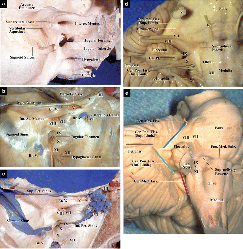
Fig. 8.1
The lateral and medial walls of the CPA. (a) Left temporal bone; posterior view. The lateral wall is formed by the temporal and occipital bones (from Matsushima T [9] with permission). (b) Left lateral wall in a specimen with the cerebellum removed. The CNs, Meckel’s cave, acoustic meatus, jugular foramen, and venous system can be observed. The bridging veins are present near the Meckel’s cave and jugular foramen (from Matsushima T [9] with permission). (c) The entire view of the venous system in the lateral wall. The dura mater covering the venous system has been removed (from Matsushima T [9] with permission). (d) Medial wall of CPA. The medial wall is formed by the brainstem and petrosal cerebellar surface. The flocculus and choroid plexus are located in the middle portion of CPA (from Matsushima T [9] with permission). (e) Fissures in CPA. The petrosal fissure and the superior and inferior limbs of the cerebellopontine fissure are observed and indicated by colored lines. They are continuous with the cerebellomesencephalic and cerebellomedullary fissures. Orange, cerebellomesencephalic fissure; blue, cerebellopontine fissure (Sup. Limb); brown, petrosal fissure; green, cerebellopontine fissure (Inf. Limb); red, cerebellomedullary fissure
The dura mater of the lateral wall contains many sinuses, including the sigmoid, superior petrosal, inferior petrosal, basilar, and marginal sinuses (Fig. 8.1c). The superior petrosal, inferior petrosal, and basilar sinuses are connected superiorly with the cavernous sinus, whereas the sigmoid and inferior petrosal sinuses are connected inferiorly with the jugular bulb, venous plexus of the hypoglossal canal, and/or posterior and lateral condylar emissary veins in the lateral foramen magnum [3, 21]. The superior petrosal veins (Sup. Pet. Vs.), which are bridging veins draining into the superior petrosal sinus from the cerebellar hemisphere, are often observed between the Meckel’s cave and upper portion of the internal acoustic meatus. The inferior petrosal veins are observed near Dorello’s canal because the inferior petrosal sinus courses around it. Small bridging veins are frequently present around the jugular foramen (Fig. 8.1b). (With regard to Sup. Pet. V., refer to Chap. 5: “The Bridging Veins in the Posterior Cranial Fossa.”)
8.3 The Medial Wall of the Cerebellopontine Angle
The medial wall of CPA is formed by the brainstem and petrosal cerebellar surface (Fig. 8.1d, e). CNs V and VII–XII originate from the lateral surface of the brainstem. The petrosal cerebellar surface is bordered superiorly from the tentorial (superior) cerebellar surface by the anterolateral margin, which corresponds to the angle formed by the petrous bone and tentorium. The anterolateral margin faces the superior petrosal sinus, which runs in the dura mater on the edge of the petrous bone. Conversely, the lower petrosal cerebellar surface gradually transits into the suboccipital (posterior) cerebellar surface, thus making the borders of these surfaces unclear.
A large fissure termed the petrosal fissure exists in the center of the petrosal cerebellar surface, the floor of which is formed by the middle cerebellar peduncle. The petrosal fissure is continuous with the cerebellopontine fissure supero- and inferomedially, which is formed by the middle cerebellar peduncle and petrosal cerebellar surface (Fig. 8.1e). The cerebellopontine fissure can be divided into two limbs: the superior and inferior limbs. The superior limb is continuous with the cerebellomesencephalic fissure superiorly and the inferior limb is continuous with the cerebellomedullary fissure (CMF) inferiorly. Arteries and veins course in these fissures.
The flocculus is situated medial or inferomedial to the petrosal fissure. It is an important intraoperative anatomical landmark while locating the origins of CN VII and the vein of the cerebellopontine fissure [15, 17–19, 24]. CNs VII and VIII, which course in the middle portion of CPA, are located slightly rostral to the flocculus when observed through the lateral suboccipital approach. The choroid plexus, protruding from the foramen of Luschka, is evident slightly inferomedial to the flocculus.
CN IV originates from the dorsal midbrain, first coursing along the tentorial edge and then entering it. The large sensory root of CN V enters the superolateral side of the pons and one or two small motor nerve rootlets, distinguishable from the sensory nerve, course just medial to the sensory root in the upper CPA. CNs VI–VIII originate from the pontomedullary sulcus in a medial to lateral order. A big concavity termed the supraolivary fossette exists at the lateral end of the pontomedullary sulcus. CNs VII and VIII originate from the lateral end of the pontomedullary sulcus at a small distance from one another, although they enter the acoustic meatus as a single nerve complex [15, 16, 18]. The origins of CNs VII and VIII are located in the supraolivary fossette, and the flocculus is situated lateral to them. They are hidden by the flocculus while viewing these structures using lateral suboccipital approach. CNs IX–XI originate in a line from the retro-olivary sulcus, the lateral margin of the olive, and enter the jugular foramen. The choroid plexus and rhomboid lip, a part of the lateral recess, are situated dorsal to CNs IX and X. CN IX usually comprises one large nerve rootlet, whereas CN X comprises several small ones. The nerve rootlets of CN XII originate from the preolivary sulcus, the medial margin of the olive, and enter the hypoglossal canals as several nerve bundles.
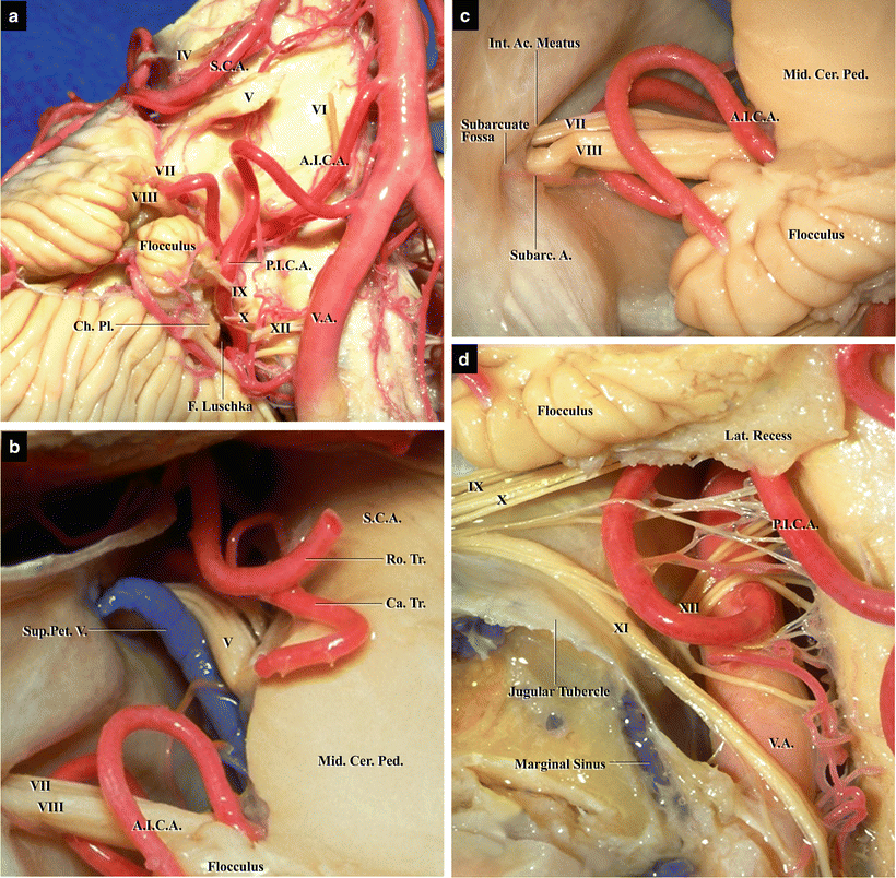
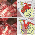
8.4 The Cranial Nerves and Cerebellar Arteries: “Rules of Three” in the Cerebellopontine Angle
Rhoton AL Jr et al. [1, 5, 6, 17] proposed the “Rules of Three in CPA,” separating each structure in CPA into three components for easy understanding of the CPA anatomy (Table 8.1). Three groups of CNs enter three bony foramina, and three cerebellar arteries intertwine with and run around them (Fig. 8.2) [17]. The three foramina, superiorly to inferiorly, are Meckel’s cave, internal acoustic meatus, and jugular foramen. Three groups of CNs, the first group comprising CN V, the second comprising CNs VII and VIII, and the third comprising CNs IX–XI, enter these three foramina. The three arteries include the superior cerebellar artery (SCA), anterior inferior cerebellar artery (AICA), and posterior inferior cerebellar artery (PICA) (Fig. 8.2).
Get Clinical Tree app for offline access
Table 8.1
“Rules of Three” in the cerebellopontine angle
Meckel’s cave—CN V—SCA—trigeminal neuralgia |
Acoustic meatus—CNs VII and VIII—AICA—hemifacial spasm |
Jugular foramen—CNs IX, X, and XI—PICA—glossopharyngeal neuralgia |
Fig. 8.2
Three cerebellar arteries and their relationships with the CNs (from Matsushima T et al. [17] with permission). (a
 The Veins of the Posterior Cranial Fossa: Nomenclature
 Three Cerebellar Arteries: Superior Cerebellar Artery, Anterior Inferior Cerebellar Artery, and Posterior Inferior Cerebellar Artery
 The Temporal Bone: Basic Anatomy and Approaches to Internal Auditory Canal
 Microvascular Decompression for Glossopharyngeal Neuralgia: Surgical Approaches Depending on the Offending Artery
 Microsurgical Anatomy of and Surgical Approaches to the Jugular Foramen
 Microsurgical Anatomy of the Cerebellomedullary Fissure and Variations of the Transcerebellomedullary Fissure Approach


Related posts:
The Veins of the Posterior Cranial Fossa: Nomenclature
Three Cerebellar Arteries: Superior Cerebellar Artery, Anterior Inferior Cerebellar Artery, and Posterior Inferior Cerebellar Artery
The Temporal Bone: Basic Anatomy and Approaches to Internal Auditory Canal
Microvascular Decompression for Glossopharyngeal Neuralgia: Surgical Approaches Depending on the Offending Artery
Microsurgical Anatomy of and Surgical Approaches to the Jugular Foramen
Microsurgical Anatomy of the Cerebellomedullary Fissure and Variations of the Transcerebellomedullary Fissure Approach
Stay updated, free articles. Join our Telegram channel
Full access? Get Clinical Tree
