(1)
Department of Neurosurgery Faculty of Medicine, Saga University, Saga, Japan
Keywords
Bridging veins in the posterior cranial fossaBridging veins on the tentorial (superior) cerebellar surfaceTentorial sinusesBridging veins on the petrosal (lateral) cerebellar surfaceSuperior petrosal veinsComplications following superior petrosal vein sacrifice5.1 Introduction
Many neurosurgeons are concerned about sacrificing the bridging veins during surgeries in the posterior cranial fossa because it occasionally causes some complications. Small bridging veins can be safely sacrificed; however, in case of large ones, it is difficult to foretell whether large bridging veins can be safely obliterated. In this chapter, bridging veins on the tentorial (superior), petrosal (lateral), and suboccipital (posterior) cerebellar surfaces are described from the perspective of surgical approaches [10]. The tentorial sinuses are also explained because they are related to bridging veins, especially on the tentorial cerebellar surface. Finally, the superior petrosal veins (Sup. Pet. Vs.), which reportedly cause serious complications when sacrificed, are described in detail along with methods of avoiding these complications.
5.2 The Tentorial Cerebellar Surface
Bridging veins that are encountered on the tentorial cerebellar surface during surgery comprise the vermian group above the inferior vermis in the midline and the hemispheric group on the tentorial hemispheric surface laterally [14, 15].
5.2.1 The Vermian (Medial) and Hemispheric (Lateral) Groups of Bridging Veins
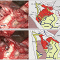
The vermian group and hemispheric group of bridging veins are also termed the medial group and the lateral group, respectively (Figs. 5.1 and 5.2a, c) [14, 15]. Vermian bridging veins, which are always located beneath the torcular herophili, are mainly formed by a collection of the terminal portions of the inferior vermian and declival veins (Figs. 5.1b and 5.2a) [14]. The vermian group includes the inferior vermian veins coursing upward in the paravermian sulci, the vein of the horizontal fissure coursing in the horizontal fissure, the declival vein coursing on the posterior part of the superior vermis, and a few tributaries in the postclival fissure.
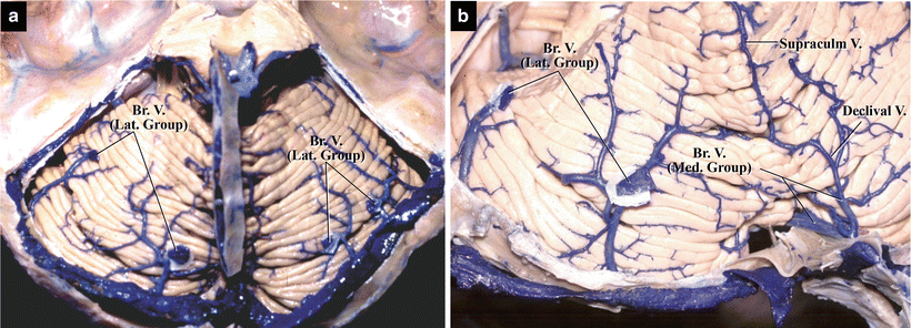
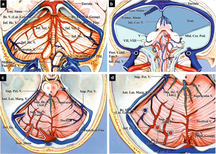
Fig. 5.1
Bridging veins on the tentorial cerebellar surface (from Matsushima T [9] with permission). (a) Hemispheric (Lateral) group. The cutting surfaces of the hemispheric group of bridging veins can be seen near the postclival fissure. (b) Vermian (Medial) group and lateral group. The vermian group of bridging veins are also visible in the midline just below the torcula
Fig. 5.2
Illustrations related to bridging veins on the tentorial cerebellar surface. (a) Their relationships to the superficial veins on the suboccipital cerebellar surface. Posterior view. The inferior hemispheric and inferior vermian veins course upward to drain into the bridging veins on the tentorial cerebellar surface (from Matsushima T et al. [14] with permission). (b) The inferior surface of the tentorium. Posterior view. The cerebellum has been removed, and the cutting surface of bridging veins on the tentorial cerebellar surface and the Group 2 tentorial sinuses can be seen (from Matsushima T [9] with permission). (c) Bridging veins and the Group 2 tentorial sinuses. Bridging veins communicate with the torcula or transverse sinus through the tentorial sinuses (from Matsushima T [14] with permission). (d) Relationships between the superficial veins and bridging veins on the tentorial cerebellar surface. Cutting surfaces of the hemispheric bridging veins can be seen near the left postclival fissure. The superior and inferior hemispheric veins join together to form the bridging veins (from Matsushima T et al. [9] with permission)
Conversely, the hemispheric group of bridging veins are formed by a collection of the terminal portions of the superior and inferior hemispheric veins and drain into the tentorial sinus (Figs. 5.1, 5.2, and 5.4) [14, 15]. Therefore, the presence and number of hemispheric bridging veins depend on the course of the superior and inferior hemispheric veins. When the inferior hemispheric veins course longitudinally upwards on the suboccipital cerebellar surface to the posterolateral margin, the hemispheric group of bridging veins usually form on the tentorial cerebellar surface. However, when the inferior hemispheric veins course diagonally in the horizontal fissure on the suboccipital cerebellar surface, the hemispheric group of bridging veins do not form on the tentorial surface (Fig. 5.2a, c). The horizontal fissure communicates between the petrosal fissure on the petrosal cerebellar surface and the upper end of the inferior vermian sulcus. There are usually one to three hemispheric groups of bridging veins near the postclival fissure on each side [14, 15]. They also comprise the origin of the Group 2 tentorial sinuses, which are described later. Therefore, a good understanding of the anatomical features of the Group 2 tentorial sinuses aids in recognizing the hemispheric group of bridging veins, which can be seen on three-dimensional computed tomography (3D CT) and cerebral angiography. Because the hemispheric group of bridging veins drains into the tentorial sinuses, classification of the tentorial sinuses, particularly Group 2, will be explained in the next section.
5.2.2 Classification of the Four Groups of Tentorial Sinuses
The tentorial sinuses are classified into four groups on the basis of their origin:
Group 1: very short tentorial sinuses on the superior surface of the tentorium near the transverse sinus, which are formed by bridging veins comprising the terminal portions of the cerebral hemispheric veins
Group 2: relatively large tentorial sinuses formed by a collection of bridging veins comprising the terminal portions of the superior and inferior cerebellar hemispheric veins on the posterior tentorial cerebellar surface
Group 3: fine tentorial sinuses originating from the tentorium itself
Group 4: tentorial sinuses formed by a bridging vein to the tentorial free edge, for example, the basal vein of Rosenthal (Fig. 5.3) [15]
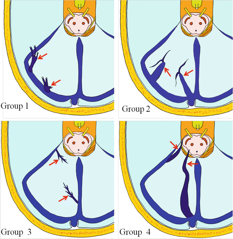
Fig. 5.3
Illustrations showing the classification of the tentorial sinuses (modified from Matsushima T et al. [15]). Group 1: The tentorial sinus receives venous blood from the cerebral hemisphere. Group 2: The sinus receives venous blood from the cerebellum. Group 3: The sinus originates from the tentorium itself. Group 4: The sinus is formed by a bridging vein to the tentorial free edge
Group 4 is often related to persistent embryonic veins. Knowledge about the Group 2 tentorial sinuses and its constituent bridging veins is critical in posterior cranial fossa surgeries, particularly those employing the infratentorial supracerebellar approach.
5.2.3 The Group 2 Tentorial Sinuses
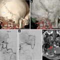
The superior hemispheric veins running downward from the anterior part of the tentorial cerebellar surface as well as the inferior hemispheric veins running upward from the posterior part of the tentorial cerebellar surface join to form bridging veins near the postclival fissure, the fissure between the simple and superior semilunar lobules situated posteriorly on the tentorial cerebellar surface. These bridging veins form the Group 2 tentorial sinuses, which drain into the torcular herophili or the transverse or straight sinuses near it (Fig. 5.4). The Group 2 tentorial sinuses can be further divided into five subtypes on the basis of their draining veins and the direction of termination (Fig. 5.5) [15]. Our previous cadaveric study revealed that the Group 2 tentorial sinuses were present in 88.5 % specimens and were always located in posterior parts of the tentorial cerebellar surface [14, 15]. The tentorial sinuses and bridging veins can be visualized before surgery using 3D CT (Fig. 5.6a); Fig. 5.6 shows 3D CT and intraoperative findings in a patient with medulloblastoma. Majority of the Group 2 tentorial sinuses drain into the torcular herophili; therefore, they can be considered similar to the tentorial draining group of the three venous draining groups of the posterior cranial fossa, according to Huang YP and Wolf BS [4].
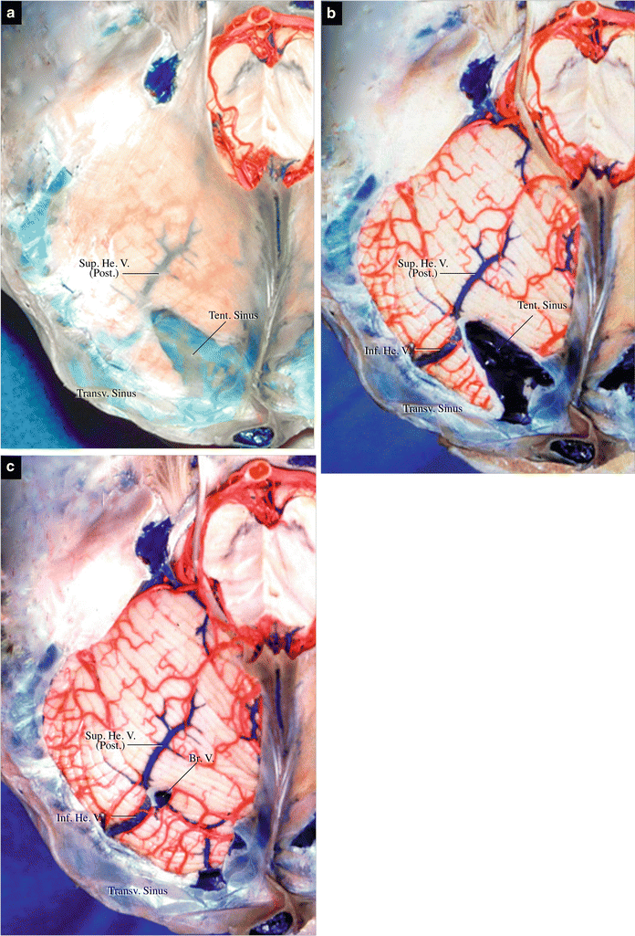
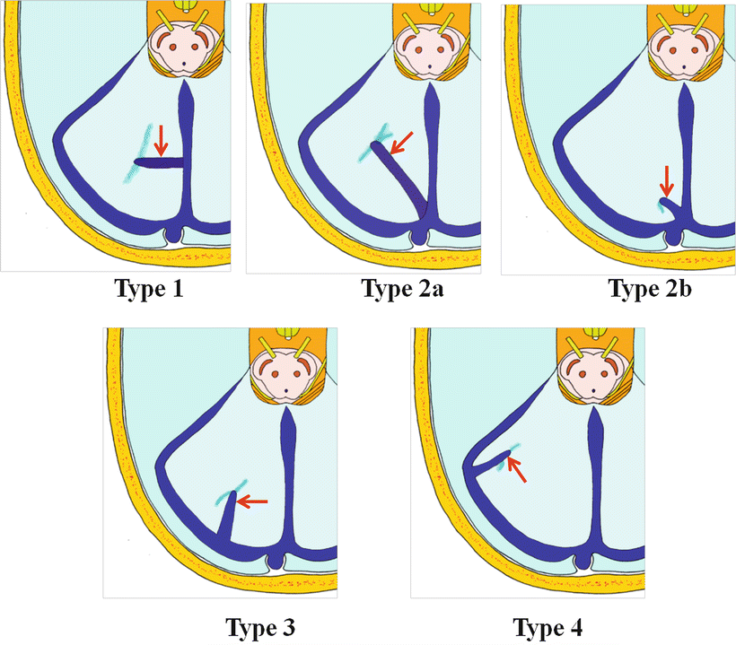
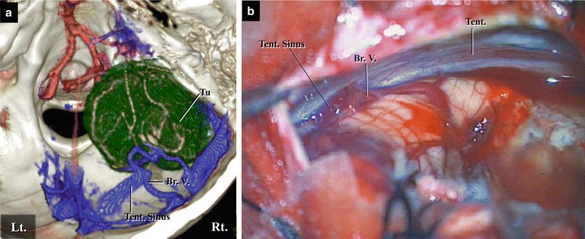
Fig. 5.4
Relationship between the bridging vein and the Group 2 tentorial sinuses (modified from Matsushima T [9]). (a) The intact tentorium. The cerebrum has been removed. (b) Group 2 of the tentorial sinus. The tentorium has been removed except for the tentorial sinus. (c) The cutting surface of bridging veins on the tentorial cerebellar surface. The tentorial sinus has been removed, and the entire tentorial cerebellar surface can be seen
Fig. 5.5
Illustrations showing the classification of the Group 2 of the tentorial sinuses (modified from Matsushima T et al. [15])
Fig. 5.6
A case of medulloblastoma in the right cerebellar hemisphere. (a) Three-dimensional computed tomography demonstrating the Group 2 tentorial sinuses and two cerebellar hemispheric veins draining into the sinuses. (b) Intraoperative photograph demonstrating bridging veins passing into the tentorial sinus after removal of the tumor
5.2.4 Bridging Veins Near the Culmen
Close to the apex of the culmen, after passing the bridging veins beneath the torcular herophili through the midline infratentorial supracerebellar approach, we encounter bridging veins near the tentorial free edge, including the superior vermian vein and/or the vein of the cerebellomesencephalic fissure. These veins converge and join to form a large vein that finally drains into the vein of Galen as a bridging vein. These veins belong to the Galenic draining group of the three draining groups of the posterior cranial fossa, according to Huang YP and Wolf BS [4]. Coagulation and incision of one of the small bridging veins, for example, the small superior vermian vein, provides a better view of the structures around the pineal body and posterior midbrain. The basal vein of Rosenthal and internal cerebral vein appear in the operative field around the pineal body; great care should be taken to avoid injuring these veins.
5.3 The Petrosal Cerebellar Surface
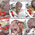
Bridging veins encountered during surgery in the cerebellopontine angle (CPA) originate from the posterior aspect of the midbrain, anterior aspect of the pons, petrosal cerebellar surface, and lateral aspect of the medulla; these veins finally drain into the superior petrosal sinus, inferior petrosal sinus, jugular bulb, marginal sinus, or sigmoid sinus (Fig. 5.7) [10, 14]. The most dangerous bridging veins are the Sup. Pet. Vs., which are often present between the upper part of the internal acoustic meatus and Meckel’s cave (Figs. 5.7, 5.8, and 5.9a
 The Veins of the Posterior Cranial Fossa: Nomenclature
 The Cerebellopontine Angle: Basic Structures and the “Rules of Three”
 Microvascular Decompression Surgery for Hemifacial Spasm: The Lateral Suboccipital Infrafloccular Approach
 Occipital Artery–Posterior Inferior Cerebellar Artery Bypass Surgery
 Microvascular Decompression for Glossopharyngeal Neuralgia: Surgical Approaches Depending on the Offending Artery
 Microsurgical Anatomy of the Cerebellomedullary Fissure and Variations of the Transcerebellomedullary Fissure Approach


Related posts:
The Veins of the Posterior Cranial Fossa: Nomenclature
The Cerebellopontine Angle: Basic Structures and the “Rules of Three”
Microvascular Decompression Surgery for Hemifacial Spasm: The Lateral Suboccipital Infrafloccular Approach
Occipital Artery–Posterior Inferior Cerebellar Artery Bypass Surgery
Microvascular Decompression for Glossopharyngeal Neuralgia: Surgical Approaches Depending on the Offending Artery
Microsurgical Anatomy of the Cerebellomedullary Fissure and Variations of the Transcerebellomedullary Fissure Approach
Stay updated, free articles. Join our Telegram channel
Full access? Get Clinical Tree
