Fig. 1.1
Percentage of age-related risk for invasive BC based on highest lifetime risk of 12 %. A threefold variation of incidence is observed in all countries of Europe (53–147 cases per 100,000) in different countries, from Belgium (147), Denmark (143), France (137), Iceland (131), the Netherlands (131), the United Kingdom (129) to Ukraine (54) and Moldova (53) [1]
Approximately 5–10 % of BCs are metastatic at diagnosis; of these patients, approximately one-fifth will survive 5 years. Depending on prognostic factors, up to 30 % of node negative and up to 70 % of node-positive BCs will relapse.
The most important risk factors include genetic predisposition, exposure to oestrogens (endogenous and exogenous) and ionising radiation, low parity and history of atypical hyperplasia. The Western-style diet, obesity and consumption of alcohol also contribute to the rising incidence of BC.
BC in males is rare, contributing about 1 % of all BCs. The major risk factors include clinical disorders carrying hormonal imbalances, radiation exposure and, in particular, a positive family history and genetic predisposition as in BRCA2 mutation (see ‘Male breast cancer’ in Sect. 19.3).
Clinical breast culture and quality of care are fundamental elements, and the outpatient clinic could have a strategic function. Despite the numerous efforts to increase awareness, so many women present late with their BC. There is some evidence that interventions delivered at an individual level can promote cancer awareness in the short term, but insufficient evidence that these might promote early presentation of cancer symptoms. Insisting on awareness is still useful in order to get more long-term results, especially for future generations.
Despite the vast improvement in the last decade in diagnostic procedures, surgical techniques and other systemic therapy offering the prospect of cure or longer life, many patients still experience significant anxiety/depression and sexual dysfunction. The objective, however unwritten, should be getting workup with the least psychological toll on the patient.
Following a diagnosis of BC, a woman finds herself in a new and unfamiliar landscape. This creates different levels of stress that vary from patient to patient and need to be addressed individually with solutions tailored to each woman’s needs. Most women will remember the information provided to them in a fragmented way. They will need space and time to process and comprehend their diagnosis, so that they can cope better psychologically with the diagnosis and treatment plan.
Breast clinics are part of breast units, specialised institutions caring for a high volume of BC patients and provided by multidisciplinary teams including at least a surgeon, a radiation oncologist, a medical oncologist, a radiologist and a pathologist – all specialised in BC. Breast clinics may have a strategic function because they are involved in the earlier steps of the breast journey. The opportunity to assess and reassure a woman from the beginning avoids worries and concerns and transmits a sense of efficacy providing the right care, at the right time, for the right person in the right way. Emotional reactions have a domino effect, and it is better to act as soon as possible, before the imagination starts to run wild.
Breast clinics could be also a starting point to start raising breast awareness as well as promoting a more generic health culture (lifestyle, eating habits, mindfulness) that could restore hope and show a ‘can-do’ attitude. The last few years have seen the growth in the number of breast clinics, breast surgeons and breast nurses. Their roles have expanded to help with the increasing workload regarding BC care. What is not clear is what would be the adequate training for such new individuals. Ideally, before they start making decisions, they should have seen hundreds of patients to cover the whole range of common and rare presentations. This is also true for trainees who cover for consultant surgeon in such clinics. Perhaps, the breast surgeons of the future will be less involved in diagnosis and more active surgically, whilst a new figure will come out, that of the outpatient breast clinic doctor.
1.1.2 The Woman’s Breast Journey
By breast journey we mean the way a woman becomes aware of having a breast problem. Several distinct conditions related to the seriousness of the disease, the individual’s ability to adapt and the prevalence of psychological factors will concur to the development of the woman’s perception of the condition on three different levels.
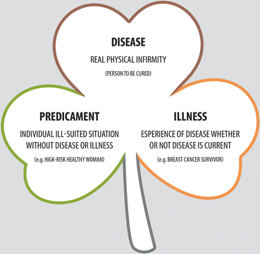
Asymptomatic healthy women, who are at high risk of cancer and ill-suited to that, have a disposition to feel they are in a predicament. Women who suffered from the disease, whether or not the disease is current, experience the condition as illness [2]. Finally, women who suffer from the actual disease experience it as such. Knowledge of the aspects of predicaments, illness and diseases could help understanding common emotional reactions observed in most women who visit breast clinics (Fig. 1.2).


Fig. 1.2
The woman’s breast journey includes perception of the risk as a predicament, disease as such, and posttreatment feelings as illness
Predicament. A predicament is a difficult situation, and it encompasses all of the psychosocial issues which impinge on the individual especially when something interrupts the regularity of their life. As a component of sickness, the predicament is more personal than the environment, diffused, multifactorial and has a very unstable structure. Predicaments can be worrisome without disease and sometimes are charged with moral and ethical implications.
Illness. An illness, too, is an experience, which may or may not be attributed to a previous disease. An illness is a social manifestation, so that an ill person with or without a disease may exhibit illness behaviour. As a component of sickness, the illness can be overplayed, stoically borne or foolishly denied. The illness is valid without a discoverable disease, and the treatment of the person with an illness is empirical and palliative.
Disease. A disease, lastly, is discernible as physical reality. Diseases are therefore amenable to scientific study and require specific changes in the structure of tissues, even though they are not necessarily organ-specific. The systematic study of a disease leads to a discovery and therefore offers scope for a specific therapy. A disease is real and valid also without illness. It may sound trivial, but a disease is real even if the changes in the body are not severe. A person with a disease should not be judged for being unable to carry out their responsibilities.
Although disease and illness are conceptually separate, the name disease is often carelessly used to describe the way someone is being ill, whilst it is important to verify whether the disease is real or if it is just experienced as such. To establish the right attitude towards the patient, the breast clinic consultant must be familiar with these conditions and be free from prejudices or pre-established assumptions. This method should be applied regardless of the psychological evaluation criteria.
1.1.3 From Primary Care to Breast Clinic
General practitioners (GPs) play a fundamental role in supporting the management of symptomatic breast patients. They should be facilitated by the specialist in their decision to refer (and to re-refer where necessary) a patient, as it is they who, in turn, will support the patient through the referral process, by providing choices and information and also through any subsequent treatment phases by providing ongoing holistic support [3].
Presentation of the patient with new breast symptoms. In the initial consultation, the GP should assess the patient with a view to refer to a symptomatic breast clinic. The GP may find that the patient has normal or benign changes that do not require referral and, at this point, he or she should give reassurance supported by the appropriate literature. All patients should be aware and informed. A good practice is not to wait for their next appointment for clinic or radiological screening if they develop symptoms.
REFERRAL TO CLINIC – Patients with the following symptoms or signs should be referred for assessment. In a patient with suspicious symptoms of BC, investigations prior to referral to a breast clinic are not recommended (expert opinion) [4].
Symptoms suggestive of urgent attention are denoted as U (urgent). Symptoms considered nonurgent but still requiring an appointment within 2 weeks are denoted as NU (not urgent). Note that family history referrals and cosmetic referrals are excluded from the 2-week wait pathway. That all patients with breast symptoms referred to a specialist should be seen within 2 weeks of referral is a quality indicator (national requirement in UK is 93 %).
Lump, lumpiness and change in texture in any woman 30 years and older that persists after next period or presents after menopause (U). The patient’s history should always be taken into account. For example, it may be appropriate, in discussion with a specialist, to agree referral within a few days also in patients reporting a lump or other symptom that has been present for several months.
Lump, lumpiness and change in texture at any age:
Discrete hard lump with fixation +/− skin tethering/dimpling/altered contour (U)
A lump that enlarges (U)
A persistent focal area of lumpiness or focal change in breast texture (U)
Progressive change in breast size with signs of oedema, local pain and reddened skin (U)
Skin distortion (U)
Previous history of BC with a new lump or suspicious symptoms (U)
Lump, lumpiness and change in texture under 30 years that do not meet the above criteria (NU). BC in women aged younger than 30 years is rare, but does occur. Benign lumps (e.g. fibroadenoma) are common, however, and a policy of referring these women urgently would not be appropriate; instead, nonurgent referral should be considered (NU).
Male patients over 50 years with unilateral firm sub-areolar mass +/− nipple discharge or associated skin changes (U).
Nipple symptoms:
Spontaneous unilateral blood-stained nipple discharge (U)
Unilateral nipple eczema or nipple change that does not respond to topical treatment (U)
Recent nipple retraction or distortion (U)
Breast Pain. Patient with minor/moderate degree of breast pain with no discrete palpable abnormality that interferes with patient’s lifestyle or sleep, when initial treatment fails, and/or with unexplained persistent symptoms (NU).
Axillary lump in the absence of clinical breast abnormality, persistent and unexplained (U).
Personal history of cancer. Patient with previously histologically confirmed BC, who presents with a further lump or suspicious symptoms, should have an urgent referral, irrespective of age (U).
High–risk woman. Request for an assessment by a patient with a strong family history of BC (NU).
WOMEN MANAGED BY GP – At least initially, GPs can manage:
Young women with tender, lumpy breast and older ones with symmetrical nodularity provided that they have no localised abnormality
Patients with minor and moderate degrees of breast pain who do not have a discrete palpable lesion
Women aged under 50 who have nipple discharge, that is, from more than one duct or is intermittent and is not blood-stained or troublesome
Aspiration of large cysts in patients with a history of multiple breast cysts, even though GPs are not commonly encouraged to do that, because bruising can follow aspiration of a solid mass, making subsequent assessment difficult
OUTCOME OF REFERRAL – Most of the patients seen in a breast clinic have normal breasts or a benign disease. Many conditions occur so commonly against the background of breast development, cyclical activity and involution that they are best considered aberrations of this process.
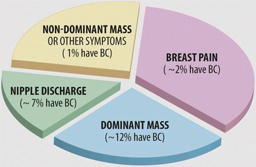
Incidence of symptoms is differently reported in single institutions. Roughly one-third have a dominant mass and one-third painful nondominant mass or lumpiness. Pain alone is referred in about 5 %, nipple discharge in 7 % and nipple retraction in 2–3 %. Rare symptoms are scaling nipple (eczema), infections and axillary lump (Fig. 1.3).


Fig. 1.3
Approximate incidence of main symptoms observed in a breast clinic
In addition, it can be observed that there are women with a strong family history of BC (see ‘High risk woman’, Sect. 2.3) or with radiological findings (as calcifications, distortion, non-palpable nodular opacities) (see Sect. 5.1).
Breast pain is common and the majority is related to the chest wall, not the breast itself (see ‘Breast pain’, Chap. 7). BC is only rarely painful, usually in older women. In young patients presenting solely with breast pain, with no palpable abnormality, there is no evidence to support the use of mammography as a discriminatory investigation for BC.
Bleeding from the nipple too is rarely due to BC. If there is a lump as well, it is more likely to be cancer. Spontaneous, single-duct persistent (>2 per week) or blood-stained nipple discharge requires a definitive diagnosis that may only be obtained by surgical duct excision (see ‘Nipple discharge’, Chap. 10).
Most benign lumps, some suspicious, usually painless and hard, are irregular in shape. A dominant breast mass can cause dimples to form in the skin of the breast or the nipple to draw in or the breast to change in size. Sometimes the skin looks like an orange peel (peau d’orange), due to lymphatic oedema around deepened hair follicles. Breast cysts diagnosed on ultrasound require aspiration only if symptomatic or complex on scan (see Sect. 9.2).
Breast infection requires early antibiotic therapy and sometimes rapid referral to hospital if it does not settle rapidly on antibiotics. Breast abscesses should be assessed by ultrasound and treated by repeated aspiration or mini-incision and drainage (see ‘Breast infections’, Sect. 8.2).
Gynaecomastia is an increasing problem. The cause should be ascertained and surgery only performed after other options have been exhausted (see ‘Gynaecomastia’ in Sect. 19.1).
Following a diagnosis of benign disease, reassurance is important, but insufficient, and an explanation of the cause, possible risks and treatment options is required. In any case, breast specialists should convey optimism about getting the control of the situation, because a patient being referred with a breast lump will be naturally concerned.
People of all ages who suspect they have BC may have particular information and support needs. The clinician should discuss these needs with the patient and respond sensitively to them. The patient should receive written and/or verbal information such as regarding accessibility of breast clinic. This information should include waiting times for an appointment and the likely process that will occur during the referral.
Primary healthcare professionals should encourage all patients, including women over 50 years old, to be breast aware in order to minimise delay in the presentation of symptoms. Patients should be reminded of the importance of keeping their appointment.
1.1.4 One-Stop Breast Clinic
A one-stop breast clinic has a theoretically considerable advantage to the formation of rapid diagnostic clinics, set up in breast units, where the diagnostic team may work together in a multidisciplinary setting [5]. Women may receive a diagnosis and management plan in the quickest time possible, in the same clinic, or have all the necessary investigations at the same time and return for results within 24–48 h.
In the case of imaging, complex investigations such as MRI, if required, may take longer to organise. In the case of pathological sampling, cytology is the only available method of obtaining an immediate definitive diagnosis, provided that it is executed by an experienced cytopathologist. Core biopsy requires a production time for pathology, and so centres that use core biopsy alone cannot currently offer a one-stop diagnostic service. Core biopsy combined with cytology is increasingly used and easier to interpret, even though partially, in case there is no conclusive result.
One-stop breast assessments are generally more favourable for people without serious disease as they go home without further waiting, knowing they do not have cancer. In fact, some patients do not require all the elements of triple assessment, as those with:
Resolved symptoms and no clinical abnormality
Clearly identified benign conditions with no other suspicious features found on clinical and imaging assessment, such as:
Areas of benign breast change and diffuse nodularity without a dominant mass
Simple cysts whether aspirated or not
Breast pain
Non-bloody nipple discharge
Gynaecomastia
Regardless of the diagnostic outcome, some patients require a long and unpredictable time to raise questions and concerns. Because the communication cannot be overcharged, closing the case in the same attendance can be counterproductive. Moreover, it is better if these more difficult patients receive information in the presence of a supportive member of their family or a friend.
The main advantages of the one-stop system are the reduction of anxiety and the possibility to provide a certain level of skill and teamwork not otherwise available. In such a service, the benefits are particularly evident for the majority of patients with normal breasts or a benign disease. Patients do like these clinics because they reduce the number of clinic visits and letters, improving administration efficiency, but surprisingly these benefits do not have long-term effects.
Although there have been concerns that immediate reporting may affect accuracy and that there may be a possible detrimental psychological aspect for those with cancer, these are more than offset by the benefits.
It is well recognised that at the time when a patient is given bad news, little other information provided in the consultation is remembered. By concentrating on establishing and delivering a diagnosis at the first visit, it is then possible to have a more useful and constructive second visit to consider the management of any cancer detected. For this reason, as previously recommended, all women with discrete masses or significant signs or symptoms must be referred directly to a specialist breast unit, and not to a basic diagnostic unit.
1.2 Landmarks of Breast Practice
Clinical Practice Points
Communication: the issue is not always about it being proper, but more about it being effective; communication is only as good as the message received.
Guidelines regarding staffing and organisation of breast units have been drawn up worldwide to ensure efficient handling of patients by appropriately experienced staff.
Guidelines fail their qualitative objectives if related to a ‘statistical’ patient. Every woman is a person who is fighting a battle you know nothing about. Being respectful is a paramount commitment. Always.
Multidisciplinary management and care are strongly recommended to avoid an opinionated standard of practice and the risk of over- or under-treat individual cases, as young or elderly or mutation carrying patients.
Especially for women who want more help with their difficulties, the breast care nurse is an essential support figure and may offer one-to-one counselling or run a support group.
The role of patients’ advocacy is also of crucial importance, particularly for the dissemination of information and knowledge.
1.2.1 Introduction
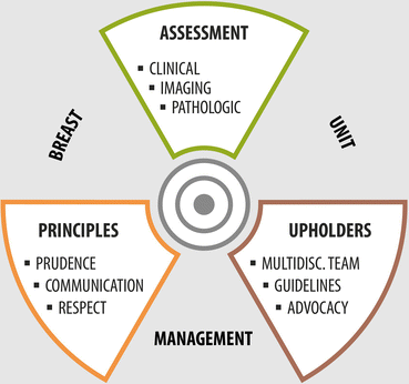
In an outpatient breast clinic, the best practice for reaching good results is likely to be founded on three ‘triples’ landmarks (Fig. 1.4).


Fig. 1.4
Landmarks of breast practice. Good practice is comprised of the triple assessment, as well interactive principles and established shields
Triple diagnostic assessment: clinical, imaging and pathologic
Triple interactive principles: communication, prudence and respect
Triple essential upholders: multidisciplinary team (MDT), evidence-based guidelines and breast care advocacy
Every workup should consider the need to fulfil all these components, which in turn are related to other issues. Outcome measures are never absolute but relative and suffer from the effects of other conditioning elements. For example, imaging is related to the age of the patient, communication is different than plane information and without respect, the slightest annoying thing can become a big concern. Guidelines fail their qualitative objectives if related to a ‘statistical’ patient.
Changing the view angle of the various elements makes the outpatient practice diversified and inspiring so that personal performance becomes worthwhile. This aspect also entails a lot of obligations but equally delivers its professional and personal rewards.
1.2.2 Triple Diagnostic Assessment (TDA)
With triple diagnostic assessment (TDA), the tests used in an individual case will be determined by the presenting symptoms, the clinical findings and the age of the patient. In some circumstances, the clinic assessment should be organised so that all appropriate tests, including imaging and needle biopsy, should be carried out during the same clinic attendance.
The breast imaging facilities should include x-ray mammography and high-frequency ultrasound. Digital mammography is preferred to film screen mammography particularly for women below 50 years and for those with dense breast tissue. Breast imaging facilities should be integrated with, or be within reasonable distance of, the breast clinic for patient convenience and efficient service delivery.
Breast MRI does not form part of the initial imaging assessment of patients in the symptomatic breast clinic. It may, however, be useful in the further investigation of some breast lesions and in the evaluation of patients with confirmed BC. In selected cases, MRI should be carried out according to the local policy agreed by the multidisciplinary team.
The TDA method will enable a diagnosis to be established in the majority of patients, and, in case of breast lumps, diagnostic surgical excision should be rarely required. The delayed diagnosis of cancers after TDA, in women who present with symptoms and are subsequently diagnosed with cancer, is approximately 0.2–0.5 %. Patients in whom the TDA is negative should be advised to seek advice from their GP if they remain concerned or if there is a change in symptoms or signs.
There should be clear administrative links between breast imaging and the breast clinic in order to ensure efficient service delivery, best use of resources, clear and rapid communication for clinic scheduling and exchange of information and results of tests.
CLINICAL ASSESSMENT – All patients who attend the symptomatic breast clinic should have a clinical consultation and physical breast examination carried out by a suitably trained member of the multidisciplinary team. This should preferably be a breast surgeon or a gynaecologist, but, in some circumstances, a radiologist, an oncologist or a trained nurse practitioner.
The consultation is aimed at establishing the nature, site and duration of the patient’s symptoms and gathering other relevant history, e.g. past history of breast disease or investigation, date of last mammogram, participation in breast screening, family history and history of HRT. It can be helpful to ask the patient to complete a questionnaire at the time of attendance at the clinic.
Minimalist clinically breast-oriented history includes:
Chief complaint, how long has it been noticed and its characteristics
Age, pregnancies, presence and regularity of menstrual periods
Personal history of breast problems and last diagnostic assessment
Family history of BC in first-degree relatives
Current taking of hormones and/or other medications
Living habits: smoking status, alcohol use, change of weight…
The physical examination (‘Clinical examination of the breast’, Chap. 4) should establish the nature and site of any abnormalities found either on visual inspection or palpation of the breast. In particular, the physical examination should establish whether there is a dominant lump present or an area of textural change. The findings of the clinical examination should be correlated with the area of concern found by the patient or referring doctor.
The physical examination should include an assessment of the axillary and supraclavicular nodes. The results of the physical examination should be recorded clearly using a diagram to indicate the site and extent of any lesions found. It is recommended that the level of physical assessment (P) should be recorded using the 1–5 scale: P1, normal; P2, benign; P3, uncertain; P4, suspicious; and P5, malignant.
IMAGING ASSESSMENT – Appropriate imaging should be carried out by suitably trained members of the multidisciplinary team, i.e. radiologist, radiographer, breast clinician, nurse and surgeon. This should preferably be a radiologist but, in some (rare) circumstances, could be a breast surgeon or a trained nurse practitioner. Ultrasound is the imaging method of choice for the majority of women aged <40 years and during pregnancy and lactation.
X-ray mammography is used in the investigation of women aged ≥40 years with the addition of ultrasound when indicated. X-ray mammography is not indicated for the majority of patients aged <40 years but should be considered in patients aged 35–39 years with clinically suspicious or malignant findings (P4, P5) and in patients with clinically indeterminate lesions (P3) if ultrasound is normal.
Stay updated, free articles. Join our Telegram channel

Full access? Get Clinical Tree


