Tendon Transfers for Management of Paralytic Deformity

CHAPTER 16 Tendon Transfers for Management of Paralytic Deformity


ANATOMY AND RELATED CONSIDERATIONS


From a simplistic perspective, any muscle (tendon) that passes anterior to the ankle joint axis functions as a dorsiflexor, and conversely, any muscle or tendon passing posterior to the axis of the ankle is a plantar flexor. This is important because the peroneal tendon and the posterior tibial tendon (PTT) are plantar flexors of the ankle, although their function is thought of primarily in terms of inversion and eversion. If a tendon lies centrally, in the axis of a joint, it exerts little influence on the motion of that joint. Conversely, the greater the distance a tendon lies from a joint axis, the greater force it exerts across the joint because of the longer lever arm. This is relevant, for example, in transfer of the PTT through the interosseous membrane. The transfer is performed subcutaneously, and the tendon is not passed inferior (deep) to the retinaculum, because this would decrease power.


The tibialis anterior muscle lies almost directly on top of the subtalar joint axis, but because it inserts on the medial cuneiform, it has an accessory function of inversion. At times, however, the tibialis anterior can become a primary invertor of the foot—for example, in the absence of a functioning tibialis posterior muscle. The Achilles tendon lies posterior to the ankle joint axis and provides the primary plantar flexion strength for the ankle. It also normally lies slightly medial to the subtalar joint axis and therefore is a weak invertor of the subtalar joint. This effect is negated with long-standing absence of the tibialis posterior muscle, in which case the peroneal tendons then pull the heel into valgus, potentiated by the valgus force of the Achilles tendon insertion. In patients with this deformity, the position of the Achilles tendon and thus the force of the gastrocnemius muscle have to be normalized by a medial translational osteotomy of the calcaneus, in addition to any tendon transfer performed.


The PTT and the peroneal tendon form a force couple around the ankle that controls hindfoot inversion and eversion. The PTT lies posterior to the ankle joint axis and medial to the subtalar axis. It therefore plantar-flexes the ankle and inverts the hindfoot, in contrast to the peroneal tendons, which plantar flex the ankle and evert the hindfoot. Paralysis of a component of this force couple allows overpull of the antagonist, resulting in varus or valgus malalignment. Although the balance of inversion and eversion may depend on the relationship between the peroneus brevis muscle and the tibialis posterior muscle, the accessory inversion of the tibialis anterior muscle and the eversion of the peroneus longus muscle (and the gastrocnemius-soleus muscle as outlined earlier) must be considered.


In planning any tendon transfer procedure, the following factors must be considered: the relative muscle strengths and tendon excursion of every functioning muscle, no matter how weak it may appear; the positioning of the tendon to be transferred relative to the rest of the foot; the proper tensioning of a transferred tendon; and the pull-out strength necessary to secure the tendon transfer. Optimally, a tendon transfer should approximate the strength and excursion of the motor unit that it is being used to replace, but such equivalent substitution can be rarely accomplished using a single tendon. Accordingly, expecting the extensor hallucis longus (EHL) muscle to replace the tibialis anterior muscle, or the flexor digitorum longus muscle to replace the tibialis posterior muscle, is unrealistic. Such a replacement can be difficult if not impossible when an attempt is made to compensate for paralysis of the strongest muscles, such as the tibialis anterior or gastrocnemius-soleus, when multiple tendon transfers may be required.


Also, it is important to consider that most muscles will lose a grade of power when transferred, particularly if the transferred tendon is not phasic (a tendon that is primarily a flexor and is transferred to function as an extensor). As an example, a PTT transfer to the dorsum of the foot to regain dorsiflexion strength is not phasic, and muscle power is lost. By contrast, if the PTT is transferred behind the ankle to the peroneal muscles to augment eversion, it is not functioning at a mechanical disadvantage because it has been kept posterior to the ankle axis. Use of a muscle that is phasic is always preferable because less “reeducation” of the muscle is required, rehabilitation is facilitated, and less strength of the muscle is lost in the transfer. Typically, in a PTT transfer for correction of a flaccid paralysis in which the tendon is passed through the interosseous membrane to the dorsum of the foot, at least one grade of muscle strength is lost. The same applies with another, nonphasic transfer such as use of the peroneal muscle(s) to substitute for absent ankle dorsiflexion. The peroneal muscle does not need to pass through the interosseous membrane (as with the PTT transfer), however, and these muscles can be passed more directly over the fibula to the anterior foot. Although this is a nonphasic transfer, less strength is lost than when a PTT transfer is used because the change in direction of the tendon transfer is minimized.


How tight should the transferred tendon be when secured to the bone? If the tendon fixed at maximal elongation, the tendon transfer serves more as a tenodesis, although it always stretches out. If it is fixed in its relaxed state, however, it cannot generate adequate tension to pull effectively. Generally, I prefer to insert the tendon under more tension than relaxation, because some stretching out of the muscle always occurs. The converse, however, does not apply, and muscle strength can never be regained if the transferred tendon is too loose. Finally, if the tendon is transferred underneath a retinaculum, which functions as a pulley, the effective tendon excursion (range of motion) is increased. With this transfer, however, the tendon is brought closer to the ankle or subtalar axes, with consequent shortening of the lever arm and reduction in strength of the transfer unit. With a subcutaneous position of a tendon transfer, excursion is decreased, but motor strength is maximized because of the greater distance from the joint axes and the resulting greater lever arm. In general, a tendon is always transferred in a subcutaneous position. Quite apart from the biomechanical advantage outlined here, the likelihood that the tendon ultimately will get “stuck,” as has been associated with transfers under the retinaculum, is greatly decreased.


Wherever possible, I perform a transfer using a tunnel with a bone-tendon-bone interference fit of the tendon. A simpler attachment of the tendon to the periosteum is never as secure. Sufficient tendon length must be present to permit its insertion in the correct location and insertion into a bone tunnel. The options for securing the tendon in the tunnel include an interference fit with a bone peg or a screw, either metallic or bioresorbable, and use of a suture anchor. Sometimes, admittedly, I use both, which ensures excellent apposition of the tendon in the tunnel, with little tendency to pull out of the bone. The fixation of the tendon is very important, because rehabilitation with weight bearing and passive range-of-motion exercises may begin once the sutures are removed, and the strengthening and retraining that need to be initiated may start sooner. Rehabilitation is essential regardless of the type of transfer, although this is easier to accomplish if the transferred tendon is in phase with the muscle it replaced.


Timing of Procedure and Preoperative Evaluation


Recovery of muscle function may occur for up to 1 year after nerve injury. An electromyogram (EMG) may have diagnostic benefit for this determination, but repeat clinical examination during this time is more helpful. Although some muscle recovery may continue up to 2 years after injury, I generally perform a transfer for paralytic deformity at 1 year after loss of function. This timing for intervention is particularly relevant when the foot is gradually deforming because of an imbalance in muscle forces about the ankle. The longer the presence of muscle imbalance, the more likely it is that fixed deformity will occur, and bone correction is required in addition to the tendon transfer. During the recovery phase after paralytic injury, the limb must be protected to prevent progressive deformity. If a protective regimen is not followed, the reconstructive procedure becomes far more difficult, if not impossible, to accomplish. A flexible equinus deformity is far easier to correct than a fixed equinovarus deformity, which may require, in addition to the tendon transfer, hindfoot and forefoot osteotomy or arthrodesis to ensure a plantigrade foot.


In evaluation of patients for possible tendon transfer, ascertaining whether the deformity is static or progressive is important. Whenever muscle imbalance is present, deformity of the foot will eventually occur, and this deterioration will be exacerbated if the muscles used for the transfer itself are involved in the paralytic process. Correction of the foot to a plantigrade position is always possible, even in a patient with a progressive deformity such as that in Charcot-Marie-Tooth disease. If the transfer is performed in childhood, however, the initial balance of the foot subsequently will be lost if the nonfunctioning muscle then strengthens. The key to an enduring result is to create a reconstruction in which the foot is both plantigrade and balanced; even if further weakening of the muscles occurs, the foot generally will remain plantigrade.


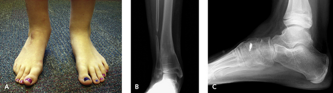
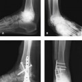
Fixed deformity of either the foot or the ankle cannot be corrected by tendon transfer alone, although the transfer may be integral to the success of surgery. For example, in a patient with a rigid equinovarus deformity, a triple arthrodesis may be chosen for correction. Although this procedure may initially correct the deformity, if tibialis posterior muscle function remains in the absence of peroneal strength (or vice versa in an equinovalgus deformity), deformity will recur, and a tendon transfer should be incorporated into the treatment plan (Figure 16-1). Any fixed deformity of the hindfoot must be corrected if a tendon transfer is performed. In order to restore passive motion across the joint on which the tendon transfer acts, the joint must be in a neutral position and the foot plantigrade. Once again, it is always preferable to use muscles that are in phase.


TECHNIQUES, TIPS, AND PITFALLS


A potential complication of a PTT transfer is a valgus deformity of the foot. This can occur in children or adults and is difficult to predict (see Figure 16-1). Although typically the result of muscle imbalance, it is also a result from incorrect positioning of the transferred tendon. Either the transfer unit needs to be moved to a more functional position or an arthrodesis should be performed.

Transfer of the PTT for correction of a paralytic equinus deformity should be performed very carefully in a patient with a preexisting flatfoot (Figure 16-2). Although the transfer is not contraindicated, a subtalar arthrodesis may need to be performed simultaneously. Another option is to transfer the PTT and simultaneously move the anterior tibial tendon under the navicular to elevate the medial foot (the same concept as that in the modified Young tenosuspension procedure for correction of flatfoot deformity). The anterior tibial tendon in this instance functions only for tenodesis and can of course stretch out.

A foot may appear to be very rigid, particularly with a cavovarus deformity, but once the PTT is transferred, the hindfoot “unwinds” (Figure 16-3). For this reason, although I may plan for an arthrodesis, until I have released the PTT and the plantar fascia, I will not commit to the procedure, because an osteotomy may be quite sufficient.

Mar 6, 2016 | Posted by in Reconstructive surgery | Comments Off on Tendon Transfers for Management of Paralytic Deformity

Full access? Get Clinical Tree

Get Clinical Tree app for offline access