Fig. 4.1
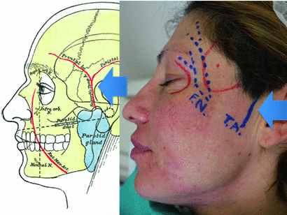
Bone anatomy of temporal fossa. Reproduction of a lithograph plate from Gray’s anatomy (Henry Gray, Anatomy: Descriptive and Surgical)
Noble structures that are contained in fossa temporalis and that need to be preserved during the treatment are as follows:
Superficial temporal artery is a major artery of the head. It arises from the external carotid artery when it bifurcates into the superficial temporal artery and maxillary artery. It begins in the parotid gland, behind the neck of the mandible, and passes superficially over the posterior root of the zygomatic process of the temporal bone; about 5 cm above this process, it divides into two branches, a frontal and a parietal. Its pulse is palpable superior to the zygomatic arch, around 1 cm anterior and superior to the tragus (Fig. 4.2, blue arrow).

Fig. 4.2
Superficial temporal artery arises from the external carotid artery. It originates in the parotid gland, behind the neck of the mandible. Its pulse is palpable superiorly to the zygomatic arch, around 1 cm anteriorly and superiorly to the tragus (blue arrow)
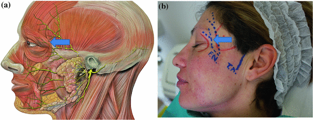
The most superior branches of the seventh cranial nerve, also known as the frontal branch of the facial nerve, cross the zygomatic arch to the temporal region, supplying the superior and anterior auricular muscles. The more anterior branches supply the frontalis, the orbicularis oculi, and corrugator supercilii. The branch crosses the zygomatic arch 3–4 cm anteriorly to the tragus and enters the temporal fossa decurring just under the auriculotemporal fascia. Accidental lesion of the temporal branch leads to facial asymmetry due to monolateral insufficient function of frontalis muscle and corrugator supercilii (Fig. 4.3a, b blue arrow).

Fig. 4.3
a, b The frontal branch of the facial nerve crosses the zygomatic arch to the temporal region, supplying the superior and anterior auricular muscles. Most of the anterior branches (blue arrow) cross the zygomatic arch 3–4 cm anteriorly to the tragus and enters the temporal fossa spreading just under the auriculotemporal fascia
4.2 Pitfalls
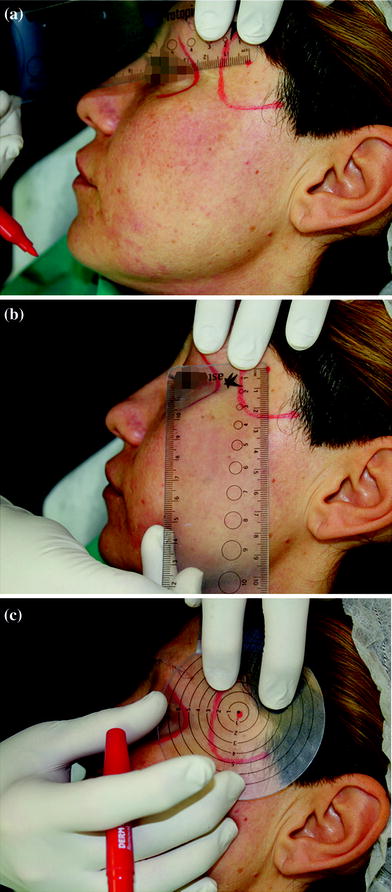
The noble structures, such as temporal artery and nerve, can be preserved using the correct point of injection (safe point). Safe point is located 2.5 cm laterally from the lateral margin of the orbital cavity (Fig. 4.4a, c) and 2.5 cm superiorly from the superior border of zygomatic arch (Fig. 4.4b, c).


Fig. 4.4
The injection is administered in a single point. Safe point of injection is located 2.5 cm laterally from the lateral margin of the orbital cavity and 2.5 cm superiorly from the superior border of zygomatic arch
The temporal artery can be touched with the finger. Its pulse can be retrieved anteriorly to the tragus, followed in the temporal area. The safe point is usually at least 2 cm anterior to this vessel (Fig. 4.4a, c).
The facial nerve is usually located at least 2 cm below the safe point (Fig. 4.4a, c).
4.3 Augmentation of the Temporal Fossa
Correction of temporal excavation can lead to substantial improvement in the appearance of the face, giving a younger look to the patient.
Augmentation of the temporal area is a simple procedure, performable in office under local anesthesia. Few noble structures need to be preserved, but this can be accomplished simply.
4.4 Augmentation of the Temporal Fossa with Calcium Hydroxylapatite
4.4.1 Indications
Marked excavation of the temporal area.
4.4.2 Contraindications
Anatomical alterations due to previous trauma or surgical intervention (neurosurgery). Scarring often induces a displacement of the noble structures and can lead to temporal artery perforation or nerve section. It can also induce retracting scars that can cause asymmetry and unequal filling of the part treated.
Stay updated, free articles. Join our Telegram channel

Full access? Get Clinical Tree


