Fig. 7.1
Lacrimal crest in red. Reproduction of a lithograph plate from Gray’s anatomy (Henry Gray, Anatomy: Descriptive and Surgical)
The eyelid is made up of several layers, from superficial to deep, which are skin, subcutaneous tissue, orbicularis oculi, orbital septum and tarsal plates, and palpebral conjunctiva.
The skin is similar to areas elsewhere, but is relatively thin and has more pigment cells. It contains sweat glands and hairs, the latter becoming eyelashes as they meet the border of the eyelid.
The orbital septum is a membranous sheet that acts as the anterior boundary of the orbit. It extends from the orbital rims to the eyelids. It forms the fibrous portion of the eyelids.
Few noble structures have to be preserved during this procedure:
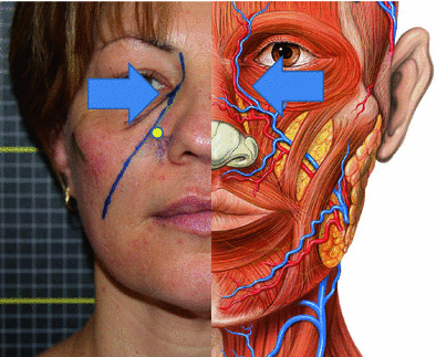
The angular artery is the terminal part of the facial artery; it ascends to the medial angle of the eye’s orbit, imbedded in the fibers of the angular head of the quadratus labii superioris, and accompanied by the angular vein (Fig. 7.2 blue arrows).

Fig. 7.2
Angular veins and arteries are indicated by the arrows. Infraorbital foramen is indicated by a yellow circle
Orbicularis Oculi: The orbicularis oculi is a muscle in the face that closes the eyelids. It arises from the nasal part of the frontal bone, from the frontal process of the maxilla in front of the lacrimal groove, and from the anterior surface and borders of a short fibrous band, the medial palpebral ligament. From this origin, the fibers are directed lateralward, forming a broad and thin layer, which occupies the eyelids or palpebrae, surrounds the circumference of the orbit, and spreads over the temple, and downward on the cheek. This muscle is thin and pale, forms a series of concentric curves, and is inserted into the lateral palpebral. The lacrimal part (tensor tarsi) is a small, thin muscle, about 6 mm in breadth, and 12 mm in length, situated behind the medial palpebral ligament and lacrimal sac. It arises from the posterior crest and adjacent part of the orbital surface of the lacrimal bone, and passing behind the lacrimal sac, divides into two slips, upper and lower, which are inserted into the superior and inferior tarsi medial to the puncta lacrimalia. The muscle acts to close the eye and is the only muscle capable of doing so.
The infraorbital nerve exits the infraorbital foramen of the maxilla and is a branch of the maxillary nerve. This nerve innervates the lower eyelid, upper lip, and part of the nasal vestibule. The infraorbital foramen (Fig. 7.2 yellow circle) is an opening in the skull located below the infraorbital margin of the orbit. It allows the passage for the infraorbital artery, vein, and nerve. The infraorbital foramen distance varies between 6.10 and 10.9 mm from the infraorbital margin. It is often palpable 1 cm below the lower orbit margin, on the pupil axis.
7.2 Pitfalls
The skin in this area is really thin, and for this reason, any irregularity of the filler injected becomes very evident. Any overcorrection with HA products in the periorbital area may cause an edematous and puffy appearance of the lower eyelids. To avoid these bad outcomes, the injections must be at a supra-periosteal level of the orbital rim: Superficial injection may lead to bad results with even a Tyndall effect. A little amount of HA should be injected: 0.05–0.1 cc of low/medium viscosity HA. Most of the times, better results can be achieved if the patient is reexamined in two weeks and then additional treatment is performed, if needed: in this area is always better a poor correction that an overcorrection with a swelling lasting for months. The HA filler should be gently massaged right after the injection to smooth down any irregularity and obtain the best distribution under the trough.
Overcorrection or irregularity with HA fillers can be corrected by off-label use of hyaluronidase.
Caution should be paid around the infraorbital foramen: It is useful to locate it with the finger before performing the pinch.
The periorbital area is prone to bruising, and in the main structures, we have to pay attention to the angular vein and arteries. The safer technique is to use a blunt-tipped cannula, diminishing in this way the chance of injuring the vessels.
7.3 Correction of Tear Trough Deformity with HA and Blunt Tip Cannule
Stay updated, free articles. Join our Telegram channel

Full access? Get Clinical Tree


