Fig. 8.1
Targets in dermal and transdermal drug delivery
Topical delivery can be defined as the application of a drug-containing formulation to the skin to directly treat cutaneous disorders or the cutaneous manifestations of a general disease. Topically delivered drugs should have their pharmacological or other effects confined to the surface of the skin or within the skin (Flynn and Weiner 1991). Formulations designed to target the skin surface include sunscreens, barrier products, cosmetics and insect repellents (Benson and Watkinson 2012). In addition to these, topical formulations can target appendages (hair follicles and sweat pores) and include antiacne products, antiperspirants, hair growth promoters and anti-infectives.
Regional delivery involves the application of a drug to the skin in order to treat diseases or alleviate disease symptoms in tissues that lie deeper, beneath the application site. Pharmacological targets of this type of drug delivery are within the musculature, vasculature, joints and tissues beneath and around the site of application. When targeting regional sites, drug formulations aim to have a regionally selective effect. Regional drug concentrations upon this route of drug administration are higher than the ones achieved by systemic administration (Flynn and Weiner 1991). For both topical and regional drug delivery, systemic absorption is unwanted but unavoidable.
In transdermal delivery drugs are applied to the skin with the aim of reaching the systemic circulation. The purpose of this type of drug delivery is to achieve a therapeutically relevant drug level in order to treat a systemic disease. Hence, the percutaneous absorption of the drug is essential, while the local deposition of the drug is unwanted, but unavoidable (Flynn and Weiner 1991). The use of transdermal delivery is limited to only a small pool of drugs (see Table 8.1) due to the selective barrier properties of the skin. The small number of candidates for this delivery route is a result of the fact that only a few drug molecules have skin permeability coefficients sufficiently high to achieve clinically active plasma levels. Currently, the market for transdermal patches comprises patches with a few low molecular weight drugs: scopolamine for motion sickness, clonidine and nitroglycerin for cardiovascular disease, fentanyl for chronic pain, nicotine to aid smoking cessation, oestradiol (alone or in combination with levonorgestrel or norethisterone) for hormone replacement and testosterone for hypogonadism (Benson 2005).
Table 8.1
List of marketed transdermal products
Generic drug | Indication | Product | Manufacturer |
|---|---|---|---|
1. Scopolamine | Motion sickness | Transderm Scop® | Novartis |
2. Nitroglycerin | Angina pectoris | Minitran®, Nitrol®, Transderm-Nitro®, Nitro-Dur® | 3 M, Rorer, Novartis, Key Pharms |
3. Clonidine | Hypertension | Catapres-TTS® | Boehringer Ingelheim |
4. Estradiol | Postmenopausal related symptoms | Estraderm®, Climara® | Novartis, Bayer HealthCare |
5. Nicotine | Smoking cessation | Nicoderm CQ®, Habitrol® | Sanofi-Aventis, Novartis |
6. Testosteron | Hypogonadism | Androderm®, Testoderm® | Watson Labs, Alza |
7. Fentanyl | Analgesia | Duragesic® | Janssen Pharmaceuticals |
8. Estradiol and levonorgestrel | Postmenopausal related symptoms | Climara ProTM | Bayer Healthcare |
9. Estradiol and norethindrone | Postmenopausal related symptoms | Combipatch® | Novartis |
10. Ethinyl estradiol and norelgestromin | Contraception | Ortho Evra® | Janssen Pharmaceuticals |
11. Buprenorphine | Analgesia | Bu Trans® | Purdue Pharma L.P. |
12. Rivastigmine | Dementia associated with Alzheimer’s and Parkinson’s disease | Exelon® | Novartis |
13. Oxybutynin | Overactive bladder | Oxytrol®, Kentera® | Watson Labs, Orion Pharma |
14. Oxybutynin chloride | Overactive bladder | Gelnique® | Watson Labs, |
15. Rotigotine | Parkinson’s disease | Neupro® | UCB Inc |
16. Granisetron | Nausea, vomiting | Sancuso® | ProStrakan Inc |
17. Methylphenidate | Attention deficithyperactivity disorder | Daytrana | Noven Pharms Inc |
18. Selegiline | Depression | Emsam® | Somerset |
19. Lidocaine | Postherpetic neuralgia pain relief | Lidoderm® | Teikoku Phar |
20. Lidocaine and tetracaine | Local dermal analgesia | Synera® | Zars Pharma |
21. Capsaicin | Postherpetic neuralgia pain relief | Qutenza® | NeurogesX |
22. Diclofenac epolamine | Topical pain relief | Flector® | Inst Biochem |
23. Diclofenac sodium | Topical pain relief in osteoarthritis | Voltaren® | Novartis |
Since percutaneous absorption is pivotal to the effectiveness of both topical and transdermal systems, significant efforts have been devoted to developing strategies to overcome the impermeability of the intact human skin. There are many ways for circumventing the stratum corneum, which provides the main barrier for drug penetration (Barry 2001).
8.3 The Skin
The skin is the largest organ in humans covering approximately 2 m2 in an average-sized adult. Its main role is to prevent water loss and protect our body from undesired outside influences. This implies that the skin acts as a barrier for diffusion of substances into the underlying tissue (Schaefer 1996; Bouwstra et al. 2003). The main problem in the dermal/transdermal administration of drugs is overcoming this natural barrier (Barry 2001; Bouwstra et al. 2003).
The skin is composed of two anatomically distinct layers: the epidermis and the dermis. Beneath the dermis is the fatty subcutaneous layer hypodermis (See Fig. 8.1). The epidermis is composed of the stratum corneum (10–20 μm thick) and the underlying viable epidermis (50–100 μm), which consists of stratum granulosum, stratum spinosum and stratum basale. The viable epidermis is responsible for the generation of the stratum corneum (Schaefer 1996).
The stratum corneum (horny layer, SC) is the final product of keratinocyte differentiation (cornification). It is made of layers of metabolically inactive cells, embedded in an extracellular matrix of lamellar lipid bilayers. Corneocytes provide the physical and chemical stability of the SC, while the extracellular matrix gives it the rigid structure, impermeable barrier for water and water-soluble compounds. The SC can be considered as a wall consisting of polyhedric squeeze-protein “bricks” and water-depleted stiff lipid sheets as “mortar” (Ghyczy 2002). The protection of the skin is provided primarily by the SC, which due to its specific structure provides the primary barrier to percutaneous absorption of compounds as well as to water loss (Lindberg and Forslind 2000; Bouwstra et al. 2003). In addition to the stratum corneum, recent findings showed that the viable epidermis is also a rate-limiting barrier to drug penetration (Andrews et al. 2012).
Corneocytes represent cell remnants of terminally differentiated keratinocytes of the viable epidermis. It is the composition of the corneocytes that gives the SC its strong barrier properties. Corneocyte layers are made up of cross-linked proteins and covalently bound lipids. The proteins resist chemical and physical denaturation and the lipids resist solubilization (Schaefer 1996; Downing and Stewart 2000; Williams 2003). In addition to these there is the corneocyte protein envelope which is added during the cornification process (Downing and Stewart 2000). The insoluble cornified envelope is stabilized through core proteins (90 % of its dry mass) cross-linked to the envelope and through covalently bound lipids (10 % of its dry mass) (Schaefer 1996). Therefore, the two layers of the envelope are the layer adjacent to the cytoplasm which is thick and composed of structural proteins and the layer on the exterior of the protein layer which is composed of lipids. The lipid layer serves as an anchor to the keratinocytes and links the proteinaceous domains to the intercellular lipid domains.
Intercellular lipids are arranged in a crystalline sublattice, with only a small portion of lipids in a liquid phase. The crystalline lipid sublattice is far less permeable to water than the liquid lipid phase. The low permeability of the SC is due not only to the unique lipid composition but also to the unique structural organization of the lipid phase (Downing and Stewart 2000; Lindberg and Forslind 2000; Bouwstra et al. 2003; Feingold et al. 1990).
The dermis (or corium) is typically 3–5 mm thick and is the major component of human skin forming the bulk of the skin. It is made of a network of connective tissue, and elastic tissue embedded in a mucopolysaccharide gel (Wilkes et al. 1973). The collagen fibres in the connective tissue give the dermis support and the elastic tissue provides flexibility. The following structures are embedded in the dermis: blood and lymphatic vessels, nerve endings, pilosebaceous units (hair follicles and sebaceous glands) and sweat glands (eccrine and apocrine). Fibroblasts, endothelilal cells and mast cells are present in the dermis, and during inflammation or wound healing, macrophages, lymphocytes and leukocytes may infiltrate (Schaefer 1996). Blood carries the molecules away from near the dermo-epidermal layer, making dermal concentrations of most permeants low. The formed concentration gradient provides the driving force for drug permeation. In addition to blood, the lymphatic system may also remove permeated molecules from the dermis, maintaining a driving force for permeation.
In terms of transdermal drug delivery, the dermis provides a minimal barrier to the delivery of most polar drugs, but may significantly limit the penetration of highly lipophilic molecules (Williams 2003).
8.4 Drug Transport Routes Through the Skin
A molecule placed on the skin surface could reach the viable tissue: (1) via the appendages, (2) across the transcellular route and (3) across the intercellular route (Fig. 8.1).
The transappendageal transport (shunt route transport) involves the transport through the pilosebaceous unit (hair follicles with sebaceous glands) and through sweat ducts. Hair follicles are the most important appendages in terms of surface area (Schaefer 1996). It is generally assumed that this pathway contributes marginally to the steady-state drug flux (Redelmeier and Kitson 1999; Agarwal et al. 2000; Barry 2006). The reason for this is that the skin area covered with the appendages is proportionally smaller than the total skin surface area (Scheuplein 1967). However, appendages may function as shunts, which may be important at short times prior to steady-state diffusion (Barry 2006). Appendages can contribute to transdermal drug delivery to a varied degree. Some results show that their (appendageal) contribution is small (Siddiqui et al. 1989), whilst others showed that these shunts are indeed important in skin permeation for a range of drugs (Illel et al. 1991). The same author (1997) also suggested that hair follicles and sebaceous glands can be privileged pathways for some molecules or formulations, which enter faster into these shunts than they do through the SC. Changing certain parameters in a formulation (such as pH, solvent, penetration enhancers) can influence follicular drug permeation (Frum et al. 2008). In addition to this some methods used for transdermal permeation enhancement, such as liposomes and iontophoresis, increase the flux of drugs through hair follicles (Li and Hoffman 1997; Hoffman 1998; Ciotti and Weiner 2002). Lauer (1999) reviewed in detail the follicular delivery.
The transcellular route leads directly across the SC, involving the drug transport through keratinocytes and intercellular lipid lamellae. The consecutive partitioning of the drug between hydrophilic (keratine) and hydrophobic (lipids) parts of the SC makes this a difficult pathway. The nature of the permeant and the partitioning coefficient will influence the importance of this route. Hydrophilic molecules may prefer the transcellular route at a pseudo-steady state. However, lipid bilayers are the rate-limiting barrier for permeation via this route (Williams 2003).
The intercellular route is through the lipid bilayers, which comprise around 1 % of the SC diffusional area, yet provide the only continuous phase within the membrane. It is generally accepted that, except for some specialized cases (e.g. highly hydrophilic substances), the intercellular lipid route is the principal pathway by which most small, uncharged molecules traverse the SC (Loth 1992; Abraham et al. 1995; Roberts et al. 1996; van Kuijk-Meuwissen et al. 1998) and many methods for enhancing the drug penetration disrupt or bypass the intercellular lipid bilayers of SC (Barry 2006). According to the domain mosaic model of the skin barrier (Forslind 1994), the structural organization of the lipids of the SC has two phases: (1) lipids in crystalline/gel state surrounded by (2) lipids that form more fluid (liquid) crystalline domains. This second, more fluid lipid domains provide the pathway by which permeants traverse the SC. A method combining ultradeformable lipid vesicles (Transfersomes®) with confocal laser scanning microscopy (CLSM) showed the existence of two different hydrophilic pathways in the SC: an “intercluster” pathway and an intercorneocyte pathway (Schätzlein and Cevc 1998). The intercluster route runs between clusters of 3–10 neighbouring corneocyte “columns”. This pathway has low resistance to molecule penetration and it comprises ≤1 % of the total skin surface or ≤20 % of the pathway area in the skin. The intercorneocyte pathway runs between all the corneocytes in a cluster and is therefore very tortuous. This transdermal permeation route resists penetration better and is more abundant (≥3 % of the skin or ≥80 % of the pathway area). Van Kuijk-Meuwissen et al. (1998) showed by CLSM that the liposomally entrapped fluorescent label travelled across SC mainly via the intercellular route.
8.4.1 Factors Affecting Drug Permeation Rate Through the Skin
Factors affecting the drug permeation rate through SC can be considered using the equation (Eq. 8.1) for steady-state flux (Barry 1983):
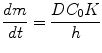
where dm/dt is the steady-state flux, presenting the cumulative mass of the diffusant, m, passing per unit area through the membrane; C 0 is the constant donor drug concentration; K is the partition coefficient of a solute between membrane and bathing solution; D is the diffusion coefficient; and h is the membrane thickness. From Eq. 8.1, the ideal properties for a molecule in order to penetrate SC well would be the following (Barry 2001; Benson 2005):
(8.1)
Low molecular mass, preferably less than 600 Da, when D tends to be high.
Adequate solubility in oil and water in order to achieve a high membrane concentration gradient, which is the driving force for diffusion (C 0 is large).
High, but balanced (optimal) K, since a too high coefficient may inhibit clearance from viable tissues. This parameter is very important in establishing a high initial penetrant concentration in the first layer of the SC. Molecules showing intermediate partition coefficients (log K octanol/water of 1–3) have adequate solubility within lipid domains of the SC (to permit diffusion through this domain) whilst still having a sufficiently hydrophilic nature to allow partitioning into the viable epidermis.
Low melting point, which correlates with good solubility as predicted by the ideal solubility theory.
When a drug possesses ideal physicochemical properties (as in the case of nicotine and nitroglycerin), transdermal delivery is feasible. If the drug does not match these ideal characteristics, manipulation of the drug or vehicle to enhance diffusion is necessary and/or penetration enhancement techniques are used.
8.5 Penetration Enhancement Technique Classification
Lots of techniques reported in literature (Barry 2001; Benson 2005; Rizwan et al. 2009) are successful in enhancing the drug delivery into/through the skin. These methods can be grouped initially into chemical and physical methods (Table 8.2). The most extensively studied chemical methods include chemical penetration enhancers (Williams and Barry 2004; Ahad et al. 2009), vesicles (El Maghraby and Williams 2009) and prodrugs (Kasting et al. 1992). Iontophoresis (Costello and Jeske 1995), electroporation (Wang et al. 1998), ultrasound (Cancel et al. 2004) and most recently microneedles (Sivamani et al. 2009) are the most studied physical methods.
Table 8.2
Methods used in transdermal penetration enhancement
Mode of action | Reference | |
|---|---|---|
Chemical enhancement methods | ||
Skin hydration | Increased drug solubility and/or disruption of the SC | Barry (2001) |
Chemical penetration enhancers | Increased drug partitioning and/or diffusion in the SC | Williams and Barry (2004) |
Vesicles | Drugs are encapsulated into vesicles which interact with the skin | Honeywell-Nguyen et al. (2004) |
Prodrugs | Chemical modification of the drug | Qandil et al. (2008) |
Ion pairs | Permeation is increased by neutralizing the drug charge with an ion of the opposite charge | Ren et al. (2008) |
Salt formation | Drug is changed into a suitable salt form to increase its solubility | Cheong and Choi (2003) |
Supersaturated solutions | Thermodynamic activity of the drug solution is shifted, thus increasing penetration rate | Dias et al. (2003) |
Eutectic systems | The mixture of drug and another substance lowers the melting point and increases solubility | Ehrenstrom and Reiz (1982) |
Physical enhancement methods | ||
Sonophoresis | Creation of microscopic holes for the transport of drugs | Tezel and Mitragotri (2003) |
Iontophoresis | Cavitational ultrasound generates shock waves that disrupt the SC lipid structure | Costello and Jeske (1995) |
Electroporation | Electrically driven transport of charged drug molecules | |
Jet injections | Pore formation with short electrical pulses | Bremseth and Pass (2001) |
Microneedles | High pressure acceleration of drug particles across the SC | Gill and Prausnitz (2007) |
Dermabrasion | Selective removal of the SC by applying high pressure microparticles | Andrews et al. (2011) |
Thermal ablation | Short intervals of localized skin heating that creates micropores | Park et al. (2008) |
Laser | Thermal ablation of SC creating pores | Gomez et al. (2008) |
Waves (radiofrequency, photomechanical, microwaves, photoacoustic) | Disruption of the structure of SC | |
Magnetophoresis | Magnetic field is driving drug movement across SC and alters the SC structure | Benson and Watkinson 2012 |
Combination of techniques | ||
Chemical enhancers and microneedles, sonophoresis and electroporation | ||
Iontophoresis and other physical methods (electroporation, sonophoresis or microneedles) | ||
Sonophoresis and other physical methods | Mitragotri et al. (2000) | |
Electroporation and microneedles | Yan et al. (2010) | |
Iontophoresis and chemical penetration enhancers | Wang et al. (2005) | |
Other methods | ||
Moxibustion | Increase in skin temperature and skin permeation | Cao et al. (2011) |
Submicron injectors | Submicron injection system isolated from sea anemone accelerates the drug across the SC | Shaoul et al. (2012) |
Mechanical methods (tape stripping, skin flexing, skin stretching, massage) | Different modes of action: removal of SC layer or reversible formation of micropathways | |
Prausnitz and Langer (2008) proposed categorizing TDDS into three generations of development. Drugs in the first generation of TDDS have low molecular weight (Mw), are lipophilic, achieve efficacy at low doses and generally do not require penetration enhancement. The second generation of TDDS utilize enhancement, such as chemical enhancers, iontophoresis and ultrasound but have been limited to the delivery of small Mw molecules. The third generation of TDDS delivers macromolecules to the SC with the help of novel chemical enhancers, electroporation, cavitational ultrasound, microneedles, thermal ablation and microdermabrasion.
8.5.1 Chemical Methods for Penetration Enhancement
Chemical penetration enhancers are defined as agents that partition into and interact with the SC constituents to induce a temporary, reversible increase in skin permeability. These substances temporarily reduce skin resistance and thereby enhance drug flux (Barry 2001). Different groups of structurally related chemical compounds are used as penetration enhancers (see Volume 3, Part 2): water, surfactants, essential oils, terpenes and their derivatives, fatty acids, esters, ethers, Azone and its derivatives, transkarbams, amides, pyrrolidones, sulphoxides and their analogues, etc. (Buyuktimkin et al. 1997; Williams and Barry 2004; Babu and Pandit 2005; Bugaj et al. 2006; Puglia and Bonina 2008; Karande and Mitragotri 2009; Mittal et al. 2009; Brychtova et al. 2010; Ibrahim and Li 2010; Karakatsani et al. 2010; Salerno et al. 2010). Chemical penetration enhancers represent the most studied penetration enhancement method as they have been shown to enhance the topical as well as transdermal delivery of a broad range of drugs both lipophilic and hydrophilic. As an example pyrrolidones enhance permeation of hydrophilic (e.g. mannitol, 5-fluorouracil and sulphaguanidine) and lipophilic drugs (betamethasone-17-benzoate, hydrocortisone and progesterone) (Williams and Barry 2004), as well as terpenes, showing enhanced skin permeation of lipophilic drugs, such as ketoprofen (Wu et al. 2001), ibuprofen (Brain et al. 2006), estradiol (Monti et al. 2002), tamoxifen (El-Kattan et al. 2001), zidovudine (Narishetty and Panchagnula 2004), hydrocortisone (El-Kattan et al. 2000) and hydrophilic drugs, e.g. propranolol hydrochloride (Zhao and Singh 1999), bupranolol (Babu and Pandit 2005), nicardipine hydrochloride (Krishnaiah et al. 2002, 2003) and others. Azone and its analogues have been used to enhance a wide range of drugs, too (Afouna et al. 2003; Jampilek and Brychtova 2012). Oleic acid is also widely studied and is one of the leading penetration enhancers used for transdermal applications (Prausnitz et al. 2004).
The limitations of using chemical enhancers are that they are not suitable for enhancing the skin penetration of high Mw drugs and that they often irritate the skin when used at concentrations necessary for achieving useful levels of penetration enhancement (i.e. they have low efficacy at low doses) (Prausnitz et al. 2004). In attempts to solve these problems, researchers have tried synthesizing novel chemical penetration enhancers (Akimoto and Nagase 2003), with optimal enhancer features such as laurocapram (Azone), which safely achieves therapeutic transport enhancement and its analogues (Jampilek and Brychtova 2012), or using two or more penetration enhancers together, because of their synergistic effect in augmenting the penetration of drugs into/through skin (Furuishi et al. 2010).
Related posts:
 The Correlation Between Transepidermal Water Loss and Percutaneous Absorption
 Epidermal Lipids and the Intercellular Pathway
 Hydrogel Vehicles for Hydrophilic Compounds
 Liposomal Gels in Enhancing Skin Delivery of Drugs
 Pickering Emulsions for Controlled Drug Delivery to the Skin
 Formulation of Drug-Cyclodextrin Complexes
Stay updated, free articles. Join our Telegram channel

Full access? Get Clinical Tree


