CHAPTER 46 Surgical Options for Femoral Reconstruction
Allograft-Prosthesis Composites
KEY POINTS
 The most important factor that determines the technique and the outcome of revision hip arthroplasty is bone stock.


Bone stock is the paramount factor that determines the appropriate technique in addressing a failing total hip arthroplasty, provides reference to the complexity of the revision procedure, and gives an indication of the expected outcome.1 The degree of bone loss is associated with the number of revision hip operations, and because younger patients are having hip arthroplasty surgery, this problem is expected to become more prevalent.
Several classification systems are used to evaluate femoral bone loss.1–3 The system that we use has five types. We have modified this system since its original publication (Table 46-1)4 and use proximal femoral allografts in types 4 and 5. With the current implant designs, we use proximal femoral allografts in femoral defects extending 8 cm or more into the femoral diaphysis. Tumor prosthesis can be used as an alternative in selected patients.
TABLE 46-1 GROSS CLASSIFICATION OF FEMORAL BONE STOCK
| Gross Classification | Type of Defect | Treatment Alternatives |
|---|---|---|
| Type 1 | No significant bone loss | Conventional cemented or uncemented femoral component |
| Type 2 | Contained (cavitary) bone loss | Proximally porous-coated implant |
| Extensively porous-coated implants for ingrowth | ||
| Extensively grit-blasted titanium implants for ongrowth | ||
| Impaction grafting with a cemented component | ||
| Modular implants for proximal or extensive ingrowth or ongrowth | ||
| Long-stemmed cemented implants | ||
| Type 3 | Segmental (full circumferential) bone loss from the proximal femur that is less than 5 cm in length and involves the calcar and the lesser trochanter but does not extend into the diaphysis | As in type 2, with a calcar replacement option |
| Type 4 | Segmental (full circumferential) bone loss of greater than 8 cm in length extending into the diaphysis | Allograft-prosthesis composite or tumor prosthesis |
| Type 5 | As in type 4, with the addition of a periprosthetic fracture | Allograft-prosthesis composite or tumor prosthesis |
INDICATIONS, CONTRAINDICATIONS, AND PITFALLS
Proximal femoral allografts in the form of allograft-prosthesis composites are indicated in hip joint reconstruction surgery either in revision total hip replacement or after tumor resection. In revision hip arthroplasty performed at our institution, full circumferential structural femoral allograft-prosthesis composites have been used in uncontained segmental femoral defects that extend for more than 8 cm into the femoral diaphysis, especially if adequate distal stabilization cannot be achieved. Another indication is the presence of Vancouver type B3 periprosthetic femoral fractures5 with significant loss of bone stock rendering the distal fixation of a long, uncemented femoral stem at least difficult, if not impossible. Proximal femoral allograft is a useful technique, particularly in young patients, because it preserves distal bone stock and potentially improves the proximal bone stock to facilitate future reconstruction.
PREOPERATIVE PLANNING
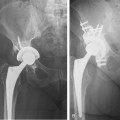
Preoperative planning will determine the level of deficient proximal femur, the approximate length and diameter of allograft required, and the correct size of the femoral component. For this purpose an anteroposterior (AP) pelvic radiograph, AP and lateral radiographs of the involved femur, and a lateral view of the involved hip are necessary. Further imaging may be required to address possible acetabulum revision issues (Fig. 46-1).
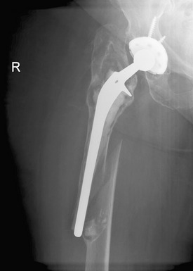
FIGURE 46-1 Preoperative radiograph. Severe osteolysis of the femur with periprosthetic femoral fracture.
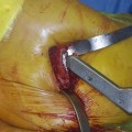
TECHNIQUE
Preparation of the Allograft
The allograft used in this procedure is stored at −70° C and in our institution is irradiated with 2.5 Mrad. Graft preparation is performed on a separate surgical table by a second surgical team while the surgical exposure is being performed by the operating surgeon. We recommend using fresh frozen allograft from an American Association of Tissue Banks–accredited tissue bank. The larger banks process the bone in bactericidal and virucidal solutions before the bone is deep frozen. Some banks provide irradiated bone, which is 10% to 20% weaker, depending on the dose.6
Stay updated, free articles. Join our Telegram channel

Full access? Get Clinical Tree


