Fig. 42.1
(a) A 9-month-old boy, extensive scald burn, debridement and mesh skin grafting. (b) A 1.5 years after surgery
In the deep dermal burns (second-degree burn, partial-thickness burns), surgical debridement is normally used together with skin grafting, but controversy is continued. Some deep dermal burn wounds may heal within 2–3 weeks, and it may not require aggressive surgical debridement of such healing wounds; however, current standard techniques and decision making of the burn depth are largely dependent on the clinicians’ naked eyes and it is often inaccurate even with experienced surgeons. A porcine contact deep dermal burn, sized 50 cm2, which requires more than 3 weeks to heal with reepithelialization by proliferation and migration of keratinocytes from the skin appendages in the deep dermis is required and this is best illustrated burn model of either with or without surgical interventions [3].
42.2.2 High-Energy Wound
As often observed in the orthopedic subspecialty, high-energy open fracture accompanies deep infection due to severe damage and remnant necrotized tissues [4]. The negative pressure wound therapy (NPWT) group developed 5.4 % of delayed deep infection, while the control did developed 28 % of deep infection. A high-energy wound is prone to wound infection due to both acute and delayed insufficiency of blood supply and severe tissue damage.
Traumatic wounds are composed of both blunt and penetrating injuries. Usually blunt trauma causes larger area of tissue damage. Crush, degloving, and avulsion high-energy injuries are in this category and the extent of the wounds is not clear all the time. There are “zone of injuries” that may be different from acute diagnosis because aggressive debridement of non-/devitalized tissues, bacterial control, and fluid maintenance may change the wound evaluation.
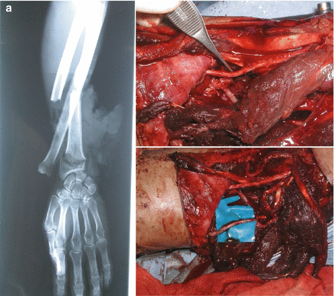
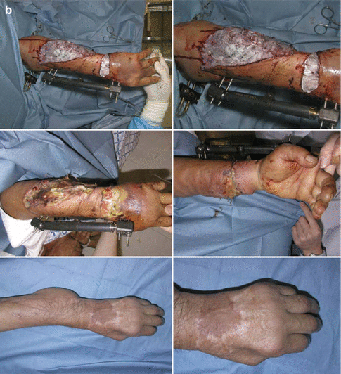
In such case, temporal coverage with artificial dermis and subsequent judgment may enhance proper healing (Fig. 42.2).


Fig. 42.2
(a) A 67-year-old man, high-energy injury, open fractures of the ulna and radius, externally intact but later found thrombus-formed radial artery (right above) and severed ulnar artery after microanastomosis. (b) External fixation with thorough debridement and artificial dermis (top), 10 days later, some tissues are still necrotic and further debridement and flap reconstruction (middle), 2 years after reconstruction (bottom)
42.2.3 Pressure Ulcer
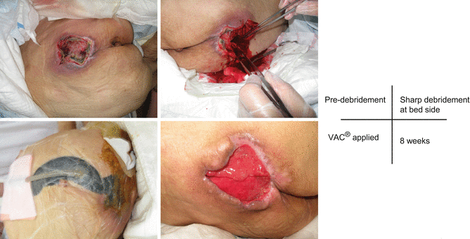
Pressure ulcers are reflecting patients’ systemic health: physical, nutritional, social, and psychological status. The complex pathophysiology suggests that several stepladders of the evolution are required. First, the sustained pressure force or shear force continued over soft tissue in between body mass, bony process, and surface. Capillary vessel flow reduced, and then blood vessel and lymphatic vessel occlusion and capillary thrombosis followed. Tissues are ischemic in this condition and reactively capillary permeability increased and the fluid is collected in the third space (extravascular space). Pressure-related intact discolored areas of the skin are described as a nonblanching erythema or suspected deep tissue injury. Prevalence of suspected deep tissue injury is more frequent than deeper pressure ulcers such as stage III or IV and increases recently than any other stages of the pressure ulcers [5]. Edematous tissues may result in necrosis, which is irreversible. Once tissue developed necrosis, precisely enough debridement is considered. The deep surgical intervention may start with when to evaluate the necrosis of tissue and how to remove effectively from the healthy surrounding tissue (Fig. 42.3).


Fig. 42.3
A 87-year-old female, sacral pressure ulcer
42.2.4 Diabetic Foot Ulcer
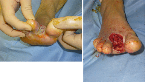
The etiology of the diabetic foot composes of combined factors including peripheral vascular disease along with sensory, motor, and autonomic neuropathy [6]. Objective evaluation for ischemia and underlying bone infection is mandatory, and surgical debridement with proper wound dressings promotes moist wound together with appropriate off-loading. Surgical debridement is considered standard of care; in theory, it may convert a chronic nonhealing wound to acute wound healing environment through deleting the senescent or non-vital cells, enhancing the wound environment to better respond to local treatment [7]. Histologic assessment from biopsies of the edge of nonhealing chronic wound demonstrates hyperproliferative epidermis with hyper- and parakeratotic elements [2] largely due to repetitive stress in the sensory-disturbed foot. Even though the keratinocytes at the nonhealing edge are activated, wound healing is deteriorated. The keratinocytes at the edge of nonhealing wound demonstrate nuclear localization of β-catenin, which leads to the downstream activation of c-Myc and glucocorticoid pathway and results in inhibition of keratinocyte migration [8]. In microarray analysis of nonhealing ulcers, reduction and cytoplasmic localization of EGFR, which causes the attenuated responsiveness to EGF, are observed. Fibroblasts from the nonhealing wound edges show clear pathogenic phenotype and slower migration [2] (Fig. 42.4).


