(1)
Department of Neurosurgery Faculty of Medicine, Saga University, Saga, Japan
Keywords
Foramen magnumFar lateral approachTranscondylar fossa approachSupracondylar transjugular tubercle approachPosterolateral approachTranscondylar approachExtreme lateral approach17.1 Introduction: A History of Skull Base Surgery in the Lateral Foramen Magnum
Approaches passing through the lateral part of the foramen magnum—the lateral foramen magnum approaches—are utilized to reach lesions at the distal vertebral artery (VA), including those at the bifurcation of VA and posterior inferior cerebellar artery (PICA), Lateral Foramen Magnum…?>on the anterolateral surface of the brainstem, or near the midline of the clivus [1–5, 10, 11, 26, 31, 33, 39–44]. These approaches have been in development since the latter half of the 1980s. Of the lateral foramen magnum approaches, the far lateral approach of Heros RC, posterolateral approach of Perneczky A, transcondylar approach of Bertalanffy H and Seeger W, and extreme lateral approach of Sen CN and Sekhar LN are the most important and widely known [5, 11, 39, 41]. Illustrations in Fig. 17.1 from their original articles show their boney openings.
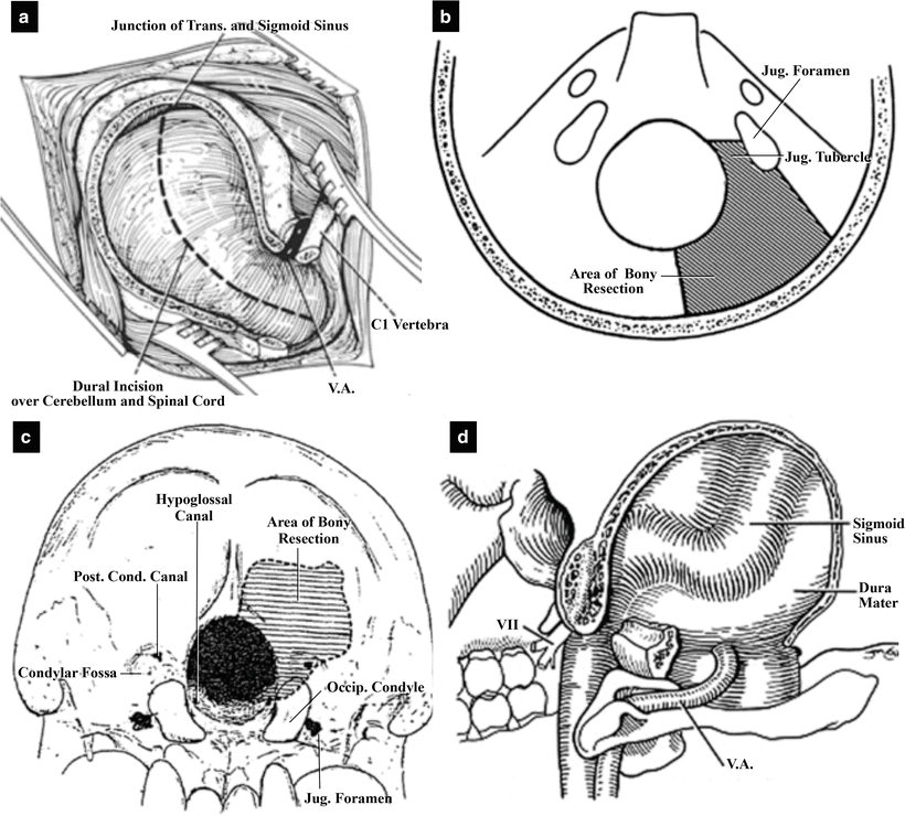
Fig. 17.1
Illustrations of the representative approaches previously reported. (a) The far lateral approach of Heros RC (from Heros RC [11] with permission). (b) The posterolateral approach to the foramen magnum of Perneczky (from Perneczky A [39] with permission). (c) The transcondylar approach of Bertalanffy H and Seeger W (from Bertalanffy H et al. [5] with permission). (d) The extreme lateral approach by Sen CN and Sekhar LN (from Sen CN et al. [41] with permission)
In 1986, Heros RC described the first lateral foramen magnum approach: the far lateral approach. It is, he stated, “a very radical removal of the bone in the area of the foramen magnum going laterally as far as the condylar fossa” [11]. However, it is difficult to define the anterior range for the removal of the condylar fossa from his description. He described the removal of the occipital bone laterally toward the sigmoid sinus, but he did not discuss the removal of the jugular tubercle (Fig. 17.1a). In the same year, Perneczky A reported the posterolateral approach to the foramen magnum [39]. He noted that drilling the jugular tubercle was particularly important and indicated the range for bony removal, which includes the jugular tubercle, in his illustration (Fig. 17.1b). His approach is unfortunately not as well known perhaps because it was introduced in a textbook. Bertalanffy H and Seeger W reported the transcondylar approach, emphasizing that they were able to remove the part of the condyle that includes the atlanto-occipital joint without any joint instability to obtain a wide operating field (Fig. 17.1c) [5]. The far lateral and transcondylar approaches had a favorable and significant influence upon surgeries in the lateral foramen magnum.
Following Bertalanffy H and Seeger W’s report, we proposed the transcondylar fossa approach (also known as the supracondylar transjugular tubercle approach) as a variation of the transcondylar approach that allows an efficient removal of bone anteriorly from the condylar fossa to the jugular tubercle without damaging the atlanto-occipital joint, and clarified the range of bony removal of the jugular tubercle as up to the hypoglossal canal (HGC) [26, 31, 33, 35]. In the approach, we suggested that the posterior condylar canal (PCC) and posterior condylar emissary vein should be used as intraoperative anatomical landmarks for accurate bony removal [23, 25, 26, 33]. The bony opening area of this approach resembles that of the posterolateral approach of Perneczky A [39]. On the other hand, it differs from the supracondylar approach reported by Gilsbach JM et al. because they did not open the foramen magnum [10]. The size of the operative field and the difficulty of the surgery vary depending on whether the foramen magnum is opened or not.
Sen CN and Sekhar LN reported the extreme lateral approach for intradural lesions of the cervical spine and foramen magnum in 1990 (Fig. 17.1d) [41]. They achieved bone exposure by removing the posterior arch of C1, performing a partial mastoidectomy, or unroofing the sigmoid sinus, depending on the longitudinal extent of the tumor. Later, Babu RP et al. reported an approach termed the extreme lateral transcondylar approach; in some cases, they mobilized VA after opening the transverse foramen of C1 [3].
Many different approaches are designated as lateral foramen magnum approaches, and it is difficult to understand their differences. They primarily differ in the area of the occipital bone removed. Salas E et al. classified the extreme lateral craniocervical approaches they experienced into six variations and presented the area of bone removed in each variation using schematic drawings [40]. The lateral foramen magnum approaches vary in whether the condyle is removed, which part of the condyle is removed, how much of the jugular tubercle is removed, whether C1 laminectomy is undertaken, and whether VA is mobilized.
The craniocervical junction is formed by C1 and the occipital bone, and the atlanto-occipital joints lie lateral to it (Fig. 17.2a, b). A jugular foramen and occipital condyle lie bilaterally. VAs run around the joints to enter the intradural region [8, 21, 24, 27, 33, 34, 37, 43]. VA and the atlanto-occipital joint are the biggest obstacles to the lateral foramen magnum approaches. We will describe the microneurosurgical anatomy of the lateral part of the foramen magnum and explain the transcondylar fossa approach, focusing on the anatomical differences between this approach and the transcondylar approach. These two approaches are basic approaches to intradural lesions through the lateral foramen magnum [5, 19, 20, 26, 28, 31, 33, 34]. We usually select one of the two approaches, depending on the height and spread of a lesion [28, 29, 32, 35, 36].
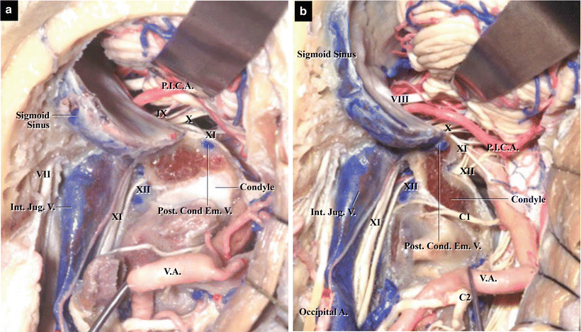
Fig. 17.2
The left lateral part of the craniocervical junction. (a) Posterior view of the sigmoid sinus and internal jugular vein, with the jugular tubercle partially removed, the vertebral artery (VA) shifted, and the cerebellum lifted. The condyle and lateral mass of C1 form the atlanto-occipital joint. The sigmoid sinus, jugular bulb, and internal jugular vein are situated in the lateral part of the foramen magnum. The posterior condylar emissary vein and lower cranial nerves (CNs) are visible. (b) Posterior view, with the condyle and jugular tubercle partially removed and the left VA transposed. The hypoglossal canal containing CN XII is visible in the center between the jugular tubercle and the condyle. The posterior condylar emissary vein communicates with the inferior end of the sigmoid sinus
17.2 Bony Structure of the Lateral Part of the Foramen Magnum
The atlanto-occipital joint is formed by the occipital condyles and the superior articular facets of the lateral mass of C1 (Fig. 17.3a, b). The lateral part of the foramen magnum comprises the jugular tubercle and occipital condyle, and HGC is located between the two (Fig. 17.3c) [8, 24, 34]. The left HGC is located at 10 o’clock, and the right HGC at the 2 o’clock position with respect to the foramen magnum [15, 16]. The jugular foramen and sigmoid sulcus are situated laterally and posterolaterally to these bony structures, respectively (Fig. 17.3c, d). When we approach via the lateral foramen magnum, the jugular tubercle and condyle form the biggest obstacles. The location and size of the condyle vary, which affects the selection of an approach. When the condyle is located more posteriorly, it often needs to be removed, whereas when it is located more anteriorly, it may be left intact.
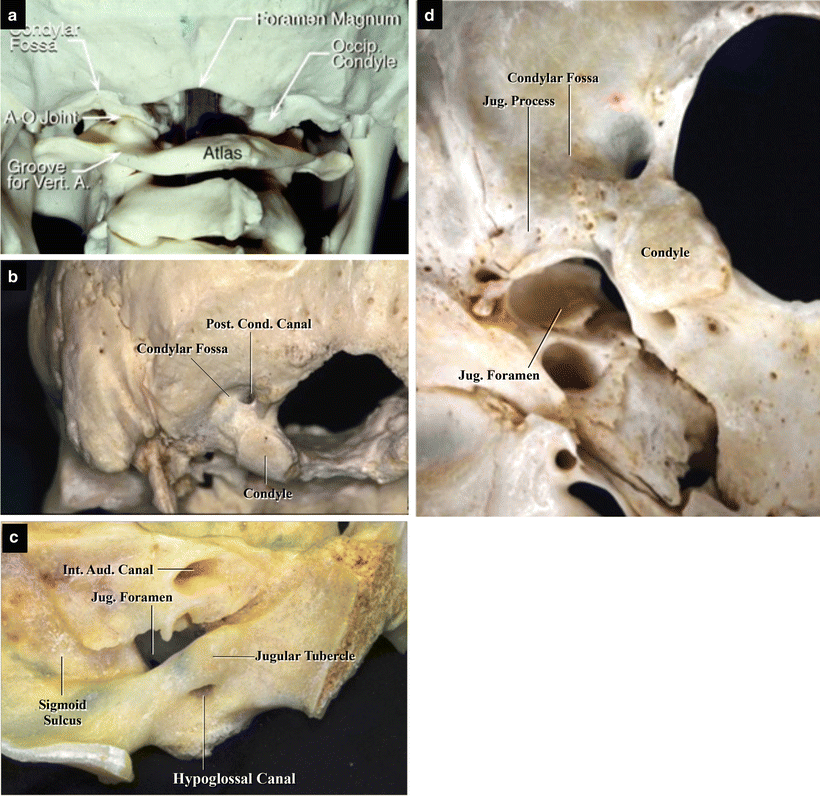
Fig. 17.3
The occipital bone and foramen magnum. (a) Posterolateral view. The paired atlanto-occipital joints and groove for the left vertebral artery are visible. The concavities behind the occipital condyles are the condylar fossae (from Matsushima T [33] with permission). (b) Posteroinferior view. The left condylar fossa is located posterior to the condyle. The posterior condylar canal can be seen at the bottom of the fossa (from Matsushima T [33] with permission). (c) Medial view. The inner surface of the left lateral wall of the foramen magnum. The left jugular tubercle and occipital condyle form the lateral part of the foramen magnum. The former is superior to the hypoglossal canal, and the latter is inferior to it. (d) Inferior view. The left jugular foramen and condylar fossa are evident lateral and posterior to the left condyle, respectively. The jugular process lateral to the condylar fossa forms the posterior wall of the jugular foramen
A hollow—the condylar fossa—lies posterosuperior to the condyle (Fig. 17.3b, d). At its base, the extracranial orifice of PCC opens, in which the posterior condylar emissary vein runs. PCC courses from the condylar fossa to the inferior end of the sigmoid sulcus posterolateral to the jugular tubercle [23, 25–27]. Then, it communicates with the posterior part of the jugular foramen or HGC anteriorly. HGC is also called the anterior condylar canal, in contrast to PCC. A venous plexus and cranial nerve (CN) XII run in the canal. The occipital bone lateral to the condylar fossa covers the sigmoid sulcus, which contains the sigmoid sinus. The jugular process lateral to the condyle forms the posterior wall of the jugular foramen (Fig. 17.3d).
17.3 Vascular Structures in the Lateral Part of the Foramen Magnum
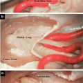
The extracranial portion of VA runs in a groove on the superior surface of the posterior arch of C1, between the occipital bone and the arch of C1, after passing through its transverse foramen (Fig. 17.4a). In its course, it runs around the atlanto-occipital joint and then passes through the dura mater to enter the intracranial region (Fig. 17.4b). VA runs inferior to the articular surface of the occipital condyle. This segment of VA gives rise to many muscular branches and is plentifully covered by the vertebral venous plexus (Fig. 17.4c). When VA penetrates the dura mater, it gives rise to the posterior spinal artery immediately before or after penetration. The intracranial segment of VA is covered with and fixed by the dentate ligament at the level of the foramen magnum (Fig. 17.4b). The nerve rootlets of CN XI cross the posterior surface of VA. When the dentate ligament is medially retracted, the C1 ventral root can be seen running ventral to VA [8, 27, 34, 43].
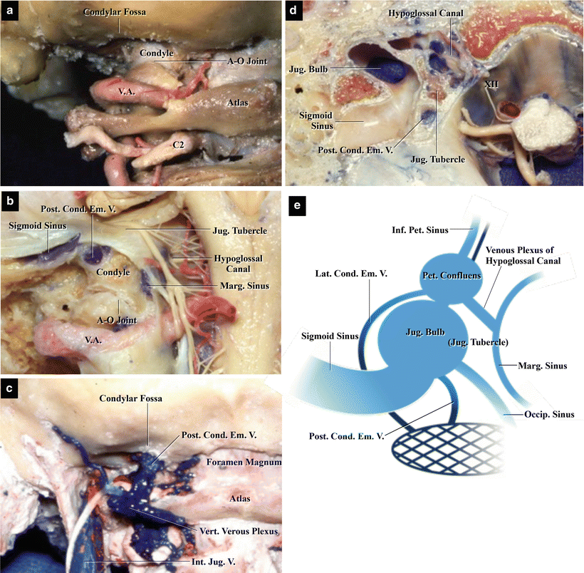
Fig. 17.4
Arterial and venous structures in the foramen magnum. (a) Posterior view with all muscles and veins removed. The extracranial portion of the left vertebral artery runs parallel to the superior margin of the left lamina of C1. It appears surrounding the atlanto-occipital joint. The left C2 nerve root can be seen below the lamina (from Matsushima T et al. [27] with permission). (b) Posterior view, with the occipital bone partially removed. The left condyle, jugular tubercle, and hypoglossal canal (HGC) are visible. The jugular tubercle is situated superior to HGC. The left posterior condylar emissary vein runs in the posterior condylar canal. The atlanto-occipital joint is situated inferior to the vein. The left sigmoid and marginal sinuses are also visible (from Matsushima T et al. [27] with permission). (c) Posterior view, with all muscles removed. The left vertebral venous plexus crosses the posterior surface of C1 and C2 and anastomoses superiorly with the left posterior condylar emissary vein (from Matsushima T et al. [27] with permission). (d) Superior view of the left lateral part of the foramen magnum. The venous system in the lateral part of the foramen magnum can be seen in the axial section at the level of HGC. The left jugular bulb, hypoglossal venous plexus, and posterior condylar emissary vein can be seen (from Matsushima T et al. [27] with permission). (e) Illustration showing the venous system in the left lateral part of the foramen magnum. There are three main draining veins from the jugular bulb, one of which is the posterior condylar emissary vein (from Matsushima K et al. [23] with permission)
There are many veins in the lateral part of the foramen magnum, and they are connected to one another. There are three main venous structures: the jugular bulb in the jugular foramen; the vertebral venous plexus surrounding the extracranial segment of VA; and the marginal sinus running in the dura mater around the foramen magnum. These venous structures are connected with one another through the posterior and lateral condylar veins and the venous plexus of HGC (Fig. 17.4b–e) [8, 37]. The posterior view of the left lateral foramen magnum presented in Fig. 17.4c shows the left posterior condylar emissary vein emerging from PCC at the inferior edge of the foramen magnum and connecting with the vertebral venous plexus. The superior view of the left lateral part of the foramen magnum presented in Fig. 17.4d demonstrates the posterior condylar emissary vein in the axial section of the jugular tubercle, venous plexus of HGC, jugular bulb, and sigmoid sinus. Figure 17.4e demonstrates the venous channels in the left lateral foramen magnum, which include three main draining points from the jugular bulb to the extracranial veins. It is crucial to understand these venous structures to perform lateral foramen magnum surgeries.
17.4 The Posterior Condylar Canal and Posterior Condylar Emissary Vein
As PCC and posterior condylar emissary vein coursing in the posterior part of the jugular tubercle represent good intraoperative anatomical landmarks when drilling during the lateral foramen magnum approaches, they will now be explained in more detail [23, 25, 31, 33]. The step-by-step dissection series presented in Fig. 17.5 shows the posterior and superior views of the left lateral foramen magnum, demonstrating the course of PCC. The extracranial orifice of PCC opens at the bottom of the condylar fossa just posterior to the condyle (Fig. 17.5a). With the occipital bone partially removed, PCC can be seen coursing in the posterior part of the jugular tubercle from behind (Fig. 17.5b). As HGC runs anterior to PCC, HGC is not exposed at this stage. HGC is exposed when the jugular tubercle is removed more anteriorly (Fig. 17.5c). The jugular tubercle is situated superior to HGC, and the condyle is inferior to it. On the resected surface of the jugular tubercle, PCC is not present anymore. In the superior view of the lateral foramen magnum, the intracranial orifice of PCC opens near the posterojugular ridge, which is the boundary between the distal end of the sigmoid sulcus and the jugular foramen (Fig. 17.5d). As reported in our previous study [23], the intracranial orifice of PCC can be classified into four types (Fig. 17.6). After removing the roof of PCC, its entire course, which runs in the posterior part of the jugular tubercle and opens at the condylar fossa, is visible (Fig. 17.5e). When the roof of HGC is partially removed, the canal can be seen anterior to the entire course of PCC. The canal is situated superior to the extracranial orifice of PCC and inferior to its intracranial orifice, in the so-called twisted position (Fig. 17.5f).
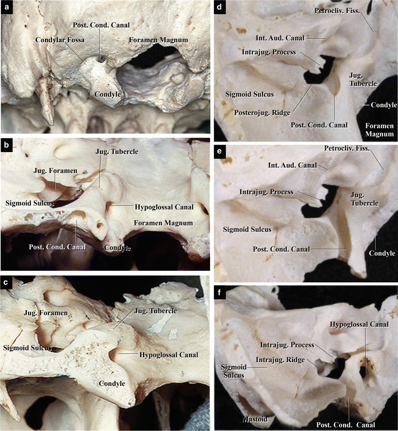
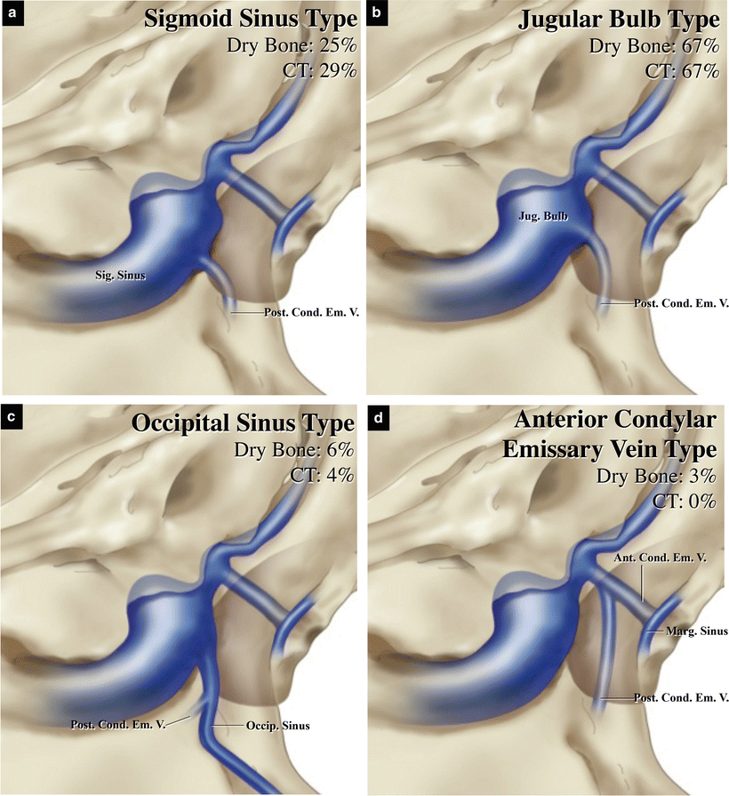
Fig. 17.5
The posterior condylar canal (PCC) in bone. Left posterior and superior views. (a) Posteroinferior view of the intact dry skull. The left condylar fossa is located posterior to the condyle. PCC opens at the bottom of the fossa. (b) Posterior view, with the occipital bone partially removed. The left PCC can be seen at the cutting surface. It courses in the posterior part of the jugular tubercle. (c) Posterior view with the posterior part of the jugular tubercle and condyle removed. The cutting surface of the left jugular tubercle and condyle is visible, but PCC is not. The jugular tubercle is superior to the hypoglossal canal (HGC) and the condyle is inferior to the canal. (d) Superior view of the intact left lateral foramen magnum. The posterojugular ridge (Posterojug. Ridge) is situated at the boundary between the distal end of the sigmoid sulcus and the jugular foramen. PCC opens in the posteromedial wall of the jugular foramen (from Matsushima K et al. [23] with permission). (e) Superior view of the occipital bone with opening of the roof of PCC. PCC runs posteroinferiorly from the jugular foramen to the bottom of the condylar fossa, making a slight internal curve (from Matsushima K et al. [23] with permission). (f) Posteromedial oblique view. The bone is slightly tilted to show HGC clearly. The medial half of the roof of HGC was also removed. The entire course of HGC, which is located anterior to PCC, can be seen. HGC runs almost horizontally between the jugular tubercle and occipital condyle (from Matsushima K et al. [23] with permission)
Fig. 17.6
Illustrations showing variation in the course of the posterior condylar emissary vein (from Matsushima K et al. [23] with permission). (a) Sigmoid sinus (SS) type. The intracranial orifice of the posterior condylar canal (PCC) opens in the posterojugular ridge, which is the boundary edge between the distal end of the sigmoid sulcus and the jugular foramen (JF). The posterior condylar emissary vein (PCEV) originates from the distal end of SS. (b) Jugular bulb (JB) type. PCC opens in the medial wall of the posterior part of JF, and PCEV originates from JB. This was the most common type in our patient cohort. (c) Occipital sinus (OS) type. The large OS descends along the posterolateral edge of the foramen magnum. PCC opens in the floor of the occipital sulcus (Occipi. Sulcus) without penetrating the occipital condyle, and PCEV originates from OS before draining into JB. (d) Anterior condylar emissary vein (ACEV) type. PCEV in PCC connects to ACEV in the hypoglossal canal intracranially
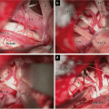
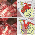
The step-by-step dissection series in Fig. 17.7 shows the left transcondylar fossa approach combined with the transcerebellomedullary fissure approach, demonstrating how PCC and posterior condylar emissary vein are considered as intraoperative anatomical landmarks, and that the condylar fossa and the jugular tubercle are obstacles.
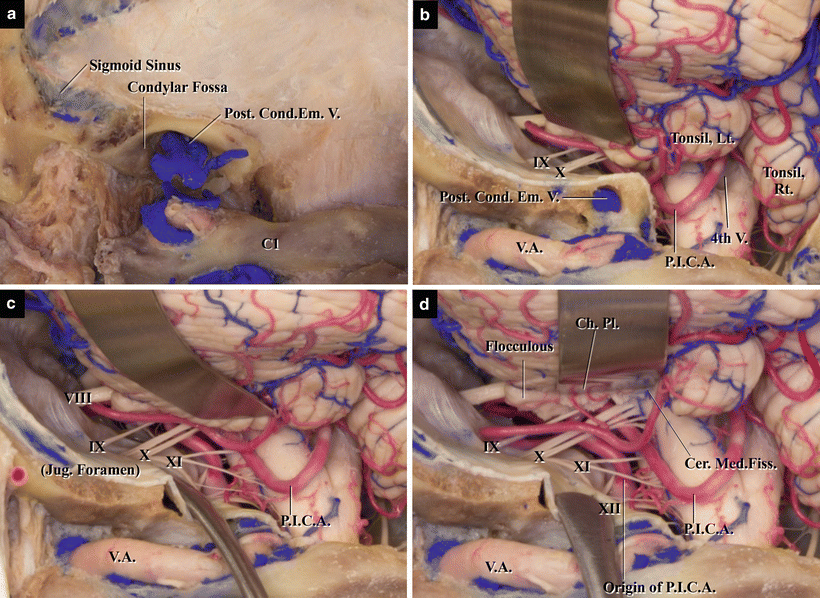
Fig. 17.7
The left transcondylar fossa approach combined with the transcerebellomedullary fissure approach in a step-by-step dissection of a cadaveric specimen. (a) Bony opening, as seen immediately after craniotomy. The condylar fossa is left intact and the large posterior condylar emissary vein is visible at its inferior aspect. (b) Dural opening. The condylar fossa is an obstacle. (c) After the removal of the condylar fossa and posterior part of the jugular tubercle, the operative field became wider. (d) The cerebellomedullary fissure has been opened
17.5 Differences Between the Transcondylar Fossa and Transcondylar Approaches
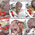
Figure 17.8a demonstrates three different approaches via the lateral part of the foramen magnum to VA–PICA aneurysms, which can originate at multiple locations of VA [32]. The VA–PICA aneurysm inferolateral to the hypoglossal canal can be treated through the midline suboccipital approach with C1 laminectomy. The approach selected depends on the level and location of an aneurysm. The main obstacles to these approaches include the atlanto-occipital joint, the occipital condyle, and the jugular tubercle. Therefore, it is necessary to understand the relationships between the extensions of bone removal and surgical view in the two basic approaches, the transcondylar fossa and transcondylar approaches.
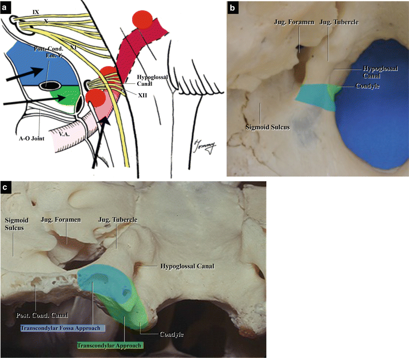
Fig. 17.8
Bony openings used in the two lateral approaches: the transcondylar fossa and transcondylar approaches. (a) Illustration demonstrating the level of each approach in cases of vertebral artery–posterior inferior cerebellar artery aneurysm. The transcondylar fossa approach is used for lesions located above the jugular foramen and hypoglossal canal (HGC), whereas the transcondylar approach is used for lesions located below HGC. (b) Drawing demonstrating the area of bone removed in each approach (blue: transcondylar fossa approach; green: transcondylar approach). Superior view. (c) Drawing demonstrating the area of bone removed in each approach (blue: transcondylar fossa approach; green: transcondylar approach). Posterior view
Figure 17.8b and c show the position of the left lateral foramen magnum in the skull, and the areas of bone removed in the two approaches are colored blue and green. The bony area removed varies depending on whether we observe superiorly or inferiorly through the operating microscope. It is necessary to remove the jugular tubercle superior to PCC to see the middle part of the clivus. To approach the anterior margin of the foramen magnum, the atlanto-occipital joint, including the condyle, must be partially removed. Recently we have opened the cerebellomedullary fissure to obtain a wide operative field for those vascular diseases as well.
PCC runs in the posterior part of the jugular tubercle toward the jugular foramen. Therefore, when PCC is followed by removing the bone superior to the canal, the transcondylar fossa approach permits partial removal of the posterior part of the jugular tubercle without damaging the atlanto-occipital joint (Fig. 17.8b, c
 The Veins of the Posterior Cranial Fossa: Nomenclature
 The Cerebellopontine Angle: Basic Structures and the “Rules of Three”
 Microvascular Decompression Surgery for Hemifacial Spasm: The Lateral Suboccipital Infrafloccular Approach
 Occipital Artery–Posterior Inferior Cerebellar Artery Bypass Surgery
 Microvascular Decompression for Glossopharyngeal Neuralgia: Surgical Approaches Depending on the Offending Artery
 Microsurgical Anatomy of the Cerebellomedullary Fissure and Variations of the Transcerebellomedullary Fissure Approach


Related posts:
The Veins of the Posterior Cranial Fossa: Nomenclature
The Cerebellopontine Angle: Basic Structures and the “Rules of Three”
Microvascular Decompression Surgery for Hemifacial Spasm: The Lateral Suboccipital Infrafloccular Approach
Occipital Artery–Posterior Inferior Cerebellar Artery Bypass Surgery
Microvascular Decompression for Glossopharyngeal Neuralgia: Surgical Approaches Depending on the Offending Artery
Microsurgical Anatomy of the Cerebellomedullary Fissure and Variations of the Transcerebellomedullary Fissure Approach
Stay updated, free articles. Join our Telegram channel
Full access? Get Clinical Tree
