Surgery for Neuropathic Foot and Ankle

CHAPTER 14 Surgery for the Neuropathic Foot and Ankle


Although classifying the Charcot arthropathic process as acute, subacute, or chronic is helpful from a practical standpoint, the definitions of these stages have no value because treatment will depend on the clinical severity of the deformity and the presence of bone fragmentation and periosteal new bone formation. New bone formation is apparent a month or so after onset of the acute process, often associated with marked osteopenia and bone fragmentation. Surgery at this stage may be more complicated even if the deformity is amenable to open reduction and internal fixation.


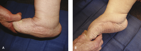
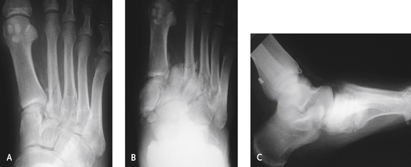
Once the process reaches the chronic phase, the midfoot typically is stable and is unlikely to deform further. However, bone prominence often is present on the plantar aspect of the midfoot, which may lead to ulceration or infection. In this context, “chronic” implies clinically stable, with an absence of swelling and inflammation (Figure 14-1). In these chronic arthropathies, the apex of the deformity is somewhere on the plantar foot. The “chronic” designation does not always equate with anatomic stability, however, because in a subgroup of midfoot arthropathies, the midfoot is quite unstable yet is not warm to touch. The deformity in such instances is very difficult to treat because of the very unstable rocker-bottom deformity of the midfoot (Figure 14-2).


Occasionally, if the first metatarsal, cuneiform, or navicular dislocates medially, the forefoot abducts and the bone prominence is directly medial. This type of deformity is easier to treat with an ostectomy than those in which bone prominences are on the lateral or plantar midfoot. The lateral rocker-bottom deformity occurs when the navicular and cuneiforms dislocate dorsally, leading to a shortening of the medial column and a laterally based prominent rocker-bottom deformity with the apex at the cuboid.


The rationale for operative treatment is to decrease the deformity, thereby minimizing the likelihood of complications, including infection and need for amputation, which otherwise may be imminent. Certainly, surgical reduction of acute dislocation makes sense, especially in patients with acute frank dislocation of the midfoot, who clearly will benefit from this more urgent operative treatment. By contrast, in patients with chronic but stable neuropathic deformity of the midfoot, use of appropriate shoes, orthoses, and braces often can restore adequate function, so the indication for surgery is more specific: deformity that cannot be controlled by nonsurgical means, associated with recurrent ulceration and infection.


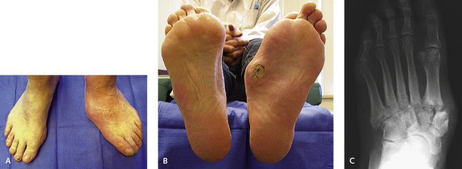
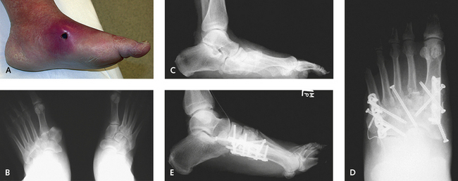
Patients can experience pain from the deformity, and although neuropathy may be thought of as being complete and not associated with any sensation, some patients experience deep, aching discomfort. Accordingly, it is appropriate to indicate surgical reconstruction with for use in such cases. Chronic deformity, recurrent ulceration, pain, and an unbraceable deformity are reasonable indications for reconstruction, but only if treatment-approach orthotics and prosthetics have failed to produce improvement or is unrealistic to begin with (Figure 14-3). There remains a “gray zone” in which the decision for performing reconstruction is more difficult if not controversial. The experienced clinician, however, gains a “feel” for the foot that helps guide decisions about the type of treatment needed. Patient factors including compliance, weight, extent of neuropathy, perfusion, skin condition, family support, opposite limb function, mobility of the ankle, and travel distance required for foot care all need to be considered in planning surgery for either the acute or the chronic stage of neuroarthropathy. The importance of these factors must not be underestimated. No matter how skillful the surgeon, the surgery will be useless if the patient lacks the means to comply with the restrictions on weight bearing and personal care requirements during rehabilitation.


CORRECTION OF NEUROPATHIC DEFORMITY OF THE MIDFOOT


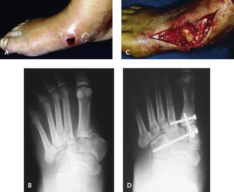
During the acute phase, some absolute indications for surgery exist, including medial dislocation of the cuneiform or navicular, which will lead to skin necrosis (Figure 14-4). Initially, the associated swelling masks the bone prominence, but with resolution of swelling, a pressure sore develops or full-thickness skin loss occurs (Figure 14-5). My experience suggests that if the dislocation is diagnosed early on in the evolution of the arthropathy, surgery will minimize the likelihood of deformity. The patient has to remain non–weight-bearing regardless of the type of treatment, and provided that the foot is not ischemic, surgery is preferable. With complete bone extrusion, operative reduction and stabilization will be necessary to prevent subsequent deformity. In patients with these acute fracture-dislocations, the issue is whether the potential morbidity of surgery outweighs the likelihood of later complications (Figure 14-6).


The key to operative treatment, however, is to take careful note of the quality of the bone. From a practical standpoint, knowing the onset of the injury is difficult, because many patients are unaware of the initial event anyway. It is preferable to use the appearance of the bone as an indicator of both the onset of the neuropathic injury and also the possibility for surgery. Performing surgery on the midfoot when the bones are crumbling as a result of osteopenia is difficult, if not frustrating, and complicated. Therefore I am more inclined to correct a subluxation or dislocation than multiple fractures around the midfoot. The traditional methods of reduction and fixation of these injuries do not work well here because they are associated with recurrence of deformity unless such techniques are combined with arthrodesis.


In the chronic stage, a complete transverse tarsal joint dislocation with dorsal dislocation of the cuneiform commonly leads to a floppy, unstable forefoot. This deformity results because the forefoot is placed into dorsiflexion by the pull of the tibialis anterior tendon while the Achilles tendon forces the hindfoot into equinus. The combination of these deformities results in a foot that is ineffective at both heel strike and toe-off, and the midfoot is at risk for ulceration.


A second deformity that frequently requires surgery in the chronic phase is the midfoot rocker-bottom deformity that is associated with supination of the forefoot. This deformity results from the heel cord pulling the foot into equinus and subsequent weight bearing on the lateral aspect of the foot.


OSTECTOMY


Ulcers may develop as a result of rubbing of anatomic bony prominences, or those produced during weight bearing in unstable joints, against shoes or other points of contact. If ulcers are intractable, ostectomy should be considered to resect the bone prominence. This procedure works well in such instances, provided that there is no associated instability of the adjacent joints. If the bone prominence is resected and the midfoot is unstable, then recurrent ulceration will occur. Ostectomy can be performed only if stability (rigidity) of the midfoot is present. Because this is a much easier and quicker procedure to recover from, with less morbidity, I prefer to perform an ostectomy, as opposed to an arthrodesis, if possible. If the ulcer fails to heal with use of a total contact cast, the ostectomy is not contraindicated. The incision has to be made carefully, however, to avoid extension of the ulcer and the possibility of infection.


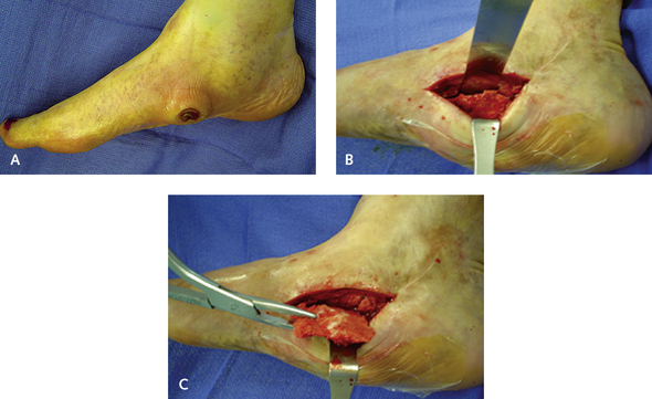
Technically, the ostectomy is not difficult to perform, and the only issue is to try to minimize postoperative soft tissue problems. Rarely, I approach the ostectomy through the open ulcer. Usually, the skin has healed over the ulcer from a total contact cast program, and the incision is made off the weight-bearing surface of the foot, either medially or laterally. Large skin flaps are preserved, and full-thickness dissection using a broad periosteal elevator should be performed to reach the prominence. I use a combination of an oscillating saw, osteotomes, and a rongeur to create a contoured surface of the plantar weight-bearing foot amenable to ambulation (Figure 14-7). It is imperative not to resect too much bone, or the result will be instability, which is particularly likely on the inferior aspect of the midfoot joints. A large, solid neuropathic bone mass may be present but is uncommonly seen; resection of the undersurface of unfused midfoot joints may have the effect of worsening the deformity and secondarily exacerbating the deformity.


TECHNIQUES, TIPS, AND PITFALLS


Stay updated, free articles. Join our Telegram channel

Mar 6, 2016 | Posted by in Reconstructive surgery | Comments Off on Surgery for Neuropathic Foot and Ankle

Full access? Get Clinical Tree

Get Clinical Tree app for offline access