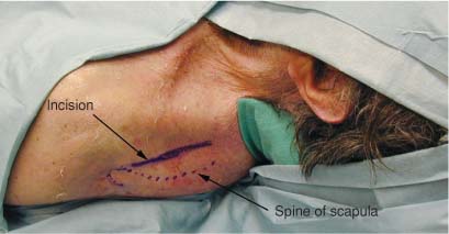
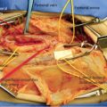
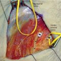
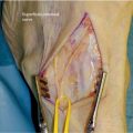
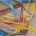
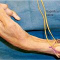
2 The suprascapular nerve arises from the upper trunk of the brachial plexus with contributions from both the C5 and C6 nerve roots. It may occasionally also receive a contribution from C4. It is the first branch off the trunk and courses lateral and deep to the trapezius muscle and enters the supraspinous fossa through the suprascapular notch, deep to the suprascapular ligament (superior transscapular ligament). The suprascapular artery travels with the nerve passing superficial to the suprascapular ligament. On occasion it may also course deep to the ligament along with the nerve. Dorsal to the notch the nerve gives off a motor ramus to the supraspinatus muscle. This ramus runs medially and penetrates the muscle on its anterior or anteroinferior aspect. The nerve then travels around the lateral border of the scapular spine, at the base of the acromion, and passes under the inferior scapular ligament (inferior transscapular ligament) together with the superior scapular vessels. Prior to entering the infraspinatous fossa, it sends small branches to the acromioclavicular joint, the subacromial bursa, and the dorsal aspect of the glenohumeral joint. Once in the infraspinatus fossa it divides into two branches that both innervate the infraspinatus muscle. The suprascapular nerve innervates the supraspinatus and infraspinatus muscles, and a small branch also supplies the suprascapular vessels. There may also be a variable dorsal ramus to the skin innervating the posterior aspect of the shoulder medial to the deltoid.1 If the proximal portion of the suprascapular nerve from its takeoff at the upper trunk of the brachial plexus is being explored, then positioning and exposure are the same for a supraclavicular brachial plexus (see Chapter 1). The supraclavicular plexus exposure may be utilized for approximately the first 10 to 12 cm of the nerve. If a more distal exposure is required, beyond the border of the trapezius, the patient is placed in the prone position. A bolster is placed under the ipsilateral shoulder to elevate it slightly off the table and allow for exposure of the upper shoulder and suprascapular area posteriorly as well as the upper portion of the supraclavicular area anteriorly. The arm is placed to the side and tucked close to the body. Elbows, wrists, knees, and ankles should be well padded to ensure no undue pressure on these bony areas. The surgeon will be working from above the patient’s head and/or to the side of the patient’s arm. An 8- to 10-cm incision is laid out midway between, and parallel to, the spine of the scapula and the clavicle. The incision should also be centered midway between the lateral edge of the shoulder and the curve of the trapezius as it swings superiorly. This placement should lie approximately in the middle of the suprascapular fossa (Fig. 2-1). The skin should be infiltrated with a vasoconstrictive agent such as lidocaine 1% with epinephrine in a 1:100,000 concentration prior to incision. After incising the skin and subcutaneous tissue, the fibers of the trapezius muscle are encountered. The trapezius muscle should be split in a medial to lateral direction (Fig. 2-2). A fatty layer overlying the supraspinatus muscle is reached (Fig. 2-3). This layer is divided exposing the supraspinatus muscle (Fig. 2-4). The supraspinatus muscle may then be carefully dissected free from the scapula subperiosteally and retracted inferiorly to expose the nerve at the suprascapular notch. The suprascapular ligament, also known as the transverse scapular ligament, crossing the notch is also visualized (Fig. 2-5). Care should be taken not to injure the suprascapular artery that traverses the notch with the suprascapular nerve (either superficial or deep to the ligament) because the subsequent bleeding will obscure the field and hemostasis is difficult to achieve in the depths of the incision. In the usual case the suprascapular artery traverses the notch superficial to the ligament. A nerve hook or a right angle clamp is placed beneath the ligament, with care being taken not to injure the nerve. The ligament is then sectioned with a blade (Fig. 2-6). The nerve may then be traced proximally as needed.
SUPRASCAPULAR NERVE
ANATOMY
POSITIONING AND SURGICAL EXPOSURE
Suprascapular Nerve
Only gold members can continue reading. Log In or Register to continue

Full access? Get Clinical Tree


