Fig. 24.1
(a–c) Preoperative view, preoperative drawings. The scar from the previous quadrantectomy is in the upper outer quadrant of the left breast. The area of the planned resection is drawn on the breast. The left breast was more ptotic than the right breast. The quadrantectomy was done as part of a superior-based pedicle reduction mammoplasty and the defect reconstructed with a de-epithelialized inferior pedicle (c)
Mammography and breast MRI found no residual microcalcifications in the breast, but due to involvement of all margins, a wide resection of the original tumor bed was planned together with a defect reconstruction (Fig. 24.1c).
24.2 Surgery
Tumor quadrantectomy was done using a superior-based pedicle reduction mammoplasty. The defect in the upper outer quadrant was reconstructed with the de-epithelialized inferior pedicle (normally being discarded in a superior pedicle reduction mammoplasty). Sentinel node biopsy revealed two negative nodes.
24.3 Clinical and Cosmetic Outcome
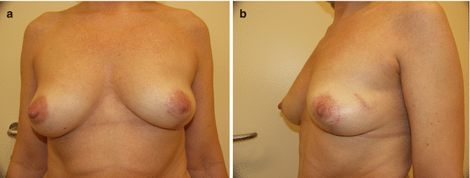
Final histology found a high-grade intraductal carcinoma in situ of a total size of 45 mm (taking into account the previous quadrantectomy) and negative margins of more than 10 mm in all directions. Postoperative radiation therapy was suggested but was declined by the patient.
The cosmetic result was rated as excellent by both the patient and the surgeon (Fig. 24.2a, b).


