Fig. 26.1
(a–d) Preoperative view, markings. The 25 mm cancer was in the upper outer quadrant of the left breast. A superior-based pedicle mammoplasty is drawn with the inferior pedicle used to reconstruct the defect (d)
26.2 Surgery
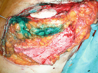
Quadrantectomy (specimen resection weight of 320 g of each breast) was performed as a superior-based pedicle reduction mammoplasty. The defect in the upper outer breast quadrant in the left breast was reconstructed with a de-epithelialized inferior pedicle normally discarded in superior-based pedicle reduction mammoplasty (Fig. 26.2). The inferior pedicle was fixed to the pectoralis major muscle with nonabsorbable sutures. Sentinel biopsy was done prior to neoadjuvant chemotherapy, and for it found two negative nodes, thus no further reoperation. Sentinel biopsy or axillary dissection was done after chemotherapy.


Fig. 26.2
Intraoperative view. The blood supply for the nipple is maintained by the superior pedicle. The tumor has already been resected, and the de-epithelialized inferior pedicle is moved up into the defect
A contralateral reduction mammoplasty using a superior-based pedicle was performed for symmetrization together with removal of the port catheter.
26.3 Clinical and Cosmetic Outcome
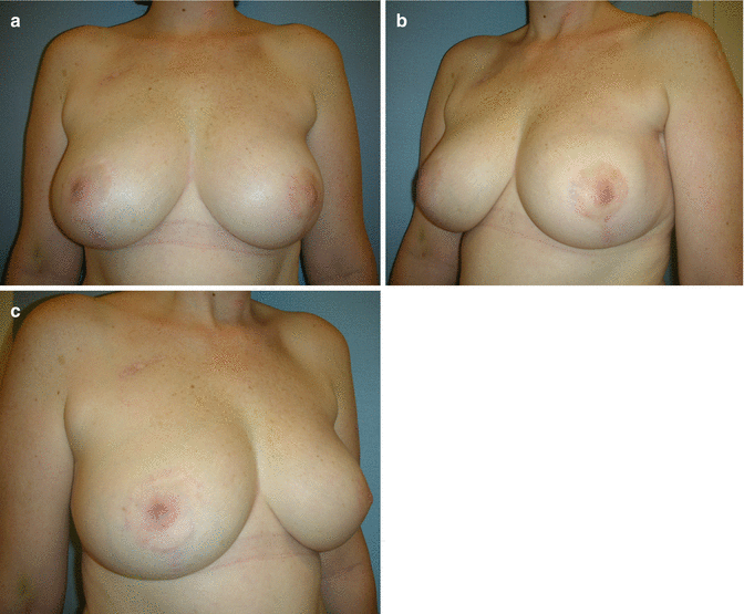
Final histology found a 15 mm tumor (yT1c) and foci of intraductal carcinoma in situ which both were completely resected with the closest margin being 17 mm in the cranial direction. The postoperative follow-up was uneventful. The patient received radiation therapy and endocrine treatment. The postoperative cosmetic result was rated as excellent by the patient and the surgeon (Fig. 26.3a–c).
Get Clinical Tree app for offline access


