Chapter 1 Structure and function of the skin
1. Name the three layers of the skin. What composes them?
The epidermis, dermis, and subcutis (subcutaneous fat). The epidermis is the outermost layer and is composed primarily of keratinocytes, or epidermal cells. Beneath the epidermis lies the dermis, which is composed primarily of collagen but also contains adnexal structures, including the hair follicles, sebaceous glands, apocrine glands, and eccrine glands. Numerous blood vessels, lymphatics, and nerves also traverse the dermis. Below the dermis lies the subcutis, or subcutaneous layer, which consists of adipose tissue, larger blood vessels, and nerves. The subcutis may also contain the base of hair follicles and sweat glands (Fig. 1-1).
2. How many layers are there in the epidermis? How are they organized?
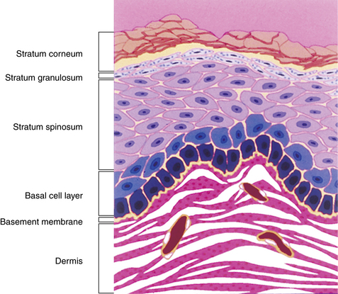
The epidermis has four layers: the basal cell layer, spiny cell layer, granular cell layer, and cornified layer (Fig. 1-2). The basal cell layer (stratum basalis) is composed of columnar or cuboidal cells that are in direct contact with the basement membrane, the structure that separates the dermis from the epidermis. The basal cell layer contains the germinative cells, and, for this reason, occasional mitoses may be present.
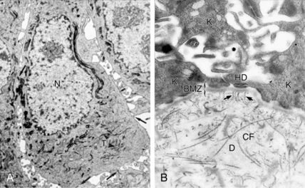
The three layers above the basal cell layer are histologically distinct and demonstrate differentiation of the keratinocytes as they move toward the skin surface and become “cornified.” Just above the basal cell layer is the spiny cell layer (stratum spinosum), so called because of a high concentration of desmosomes and keratin filaments that give the cells a characteristic “spiny” appearance (Fig. 1-3A). Above the spiny layer is the granular cell layer (stratum granulosum). In this layer, keratohyalin granules are formed and bind to the keratin filaments (tonofilaments) to form large electron-dense masses within the cytoplasm that give this layer its “granular” appearance.
3. Do other types of cells normally occur in the epidermis?
In addition to keratinocytes, three other cells are normally found in the epidermis. The melanocyte, the most common, is a dendritic cell situated in the basal cell layer. There are approximately 36 keratinocytes for each melanocyte. This cell’s function is to synthesize and secrete melanin-containing organelles called melanosomes.
5. Describe the structure of the basement membrane zone (BMZ).
The BMZ is not normally visible by light microscopy in sections stained with hematoxylin-eosin but can be visualized as a homogeneous band measuring 0.5 to 1.0 mm thick on periodic acid–Schiff staining. Ultrastructural studies and immunologic mapping demonstrate that the BMZ is an extremely complex structure consisting of many components that function to attach the basal cell layer to the dermis (Fig. 1-3B). Uppermost in the BMZ are the cytoplasmic tonofilaments of the basal cells, which attach to the basal plasma membrane of the cells at the hemidesmosome. The hemidesmosome is attached to the lamina lucida and lamina densa of the BMZ via anchoring filaments. The BMZ, in turn, is anchored to the dermis by anchoring fibrils that intercalate among the collagen fibers of the dermis and secure the BMZ to the dermis. The importance of these structures in maintaining skin integrity is demonstrated by diseases such as epidermolysis bullosa, in which they are congenitally missing or damaged.
6. How is the structure of the epidermis related to its functions?
The three most important functions of the epidermis are protection from environmental insult (barrier function), prevention of desiccation, and immune surveillance. The stratum corneum is an especially important cutaneous barrier that protects the body from toxins and desiccation. Although many toxins are nonpolar compounds that can move relatively easily through the lipid-rich intracellular spaces of the cornified layer, the tortuous route among cells in this layer and the layers below effectively forms a barrier to environmental toxins. Ultraviolet light, another environmental source of damage to living cells, is effectively blocked in the stratum corneum and the melanosomes. The melanosomes are concentrated above the nucleus of the keratinocytes in an umbrella-like fashion, providing photoprotection for both epidermal nuclear DNA and the dermis.
7. What structural components of the epidermis are involved in blistering diseases?
In the epidermis, the keratinocytes are bound to each other by desmosomal complex. These complexes consist of the desmosome and the tonofilaments of the cytoplasm that are made of cytokeratins. In the upper stratum spinosum, the tonofilaments are composed mainly of keratins 1 and 10. Congenital abnormalities in these keratins produce structural weakness between keratinocytes, producing a disease called congenital bullous ichthyosiform erythroderma


Stay updated, free articles. Join our Telegram channel

Full access? Get Clinical Tree


