Fig. 40.1
STONEFISH – Olivier Allart from Diony-Bulles (Reunion Island) All the other photos are from myself ; we are not obliged to mention the author either: (photo jdh)
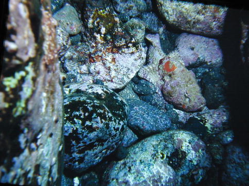
Fig. 40.2
Do you see the stonefish???
40.1.2 Habitat
Stonefishes are found on coral reefs, muddy waters or sandblasters of the Indian Ocean, Red Sea, Indonesia, Australia, New Caledonia and the Pacific [5].
40.1.3 Venomous Apparatus
40.1.3.1 Thorns and Glands with Venom
The stonefish has 13 thorns on its dorsal fin which rise in case of danger. Every thorn is covered with a verrucose tegumental envelope and has a pair of glands with venom in its base. Every gland has a canal filled with poison and leading to the top of the thorn. If someone walks on or tries to seize the stonefish, its thorns penetrate into the skin and the tegumental envelope is then pushed downward causing the compression of the gland, which if sufficient will launch the release of the poison. The stinging and poisoning is a defensive mechanism.
The venom: its composition is complex: substances such as the histamine, adrenalin, norepinephrine, dopamine as well as enzymes such as hyaluronidase, protease and lipase. The main toxin is the verrucotoxin: it inhibits the calcic canals and it activates the potassium canals. The venom is myotoxic, neurotoxic and haemolytic. It is inactivated in 54 °C [3].
40.2 Stings by Stonefish
40.2.1 General ideas
Stonefishes were notorious for being among the most venomous animals of the world and its stings were thought to be lethal. In fact, the sting by stonefish provokes an extremely intense pain that can possibly result in a faint fit. Also if it happens to a scuba diver, it may cause an accident if the diver is unable to control his ascent (barotraumatism) or to make the required decompression stops (desaturation accident).
40.2.2 Circumstances
The stonefish is a sedentary and benthic fish. It favours rocky areas or lays buried under sand or mud. It feeds on small fishes and shrimps which it swallows with its mouth. The stonefish can survive several hours out of the water and its venom remains active from 24 to 48 h after its death. Stings by stonefishes mostly occur during activities such as bathing, fishing, snorkelling and scuba diving.
40.2.3 Wound Location
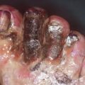
The wounds are mainly seen on the feet of people who accidentally walked on a stonefish. They can also be seen on hands due to an attempt to touch or seize the fish.
40.2.4 Clinical Evidence
The pain is immediate and very intense. It can cause a person to faint. The pain is quoted between 8 and 10 on the visual analogic pain scale. It often results in restlessness and aggressiveness.
There are one or several wounds, mostly in the foot or in the hand.
An oedema quickly develops, sometimes limited around the wound, often wider and spreading. Not rarely, it might extend to the whole limb up to its base.
General symptoms are often noticed: sweats, nauseas, arterial hypotension, tachycardia, heart rhythm disorders, myasthenia and pulmonary oedema.
40.2.5 Diagnosis
The diagnosis is easy when the victim saw the fish and clearly identified it. It is often the case with fishermen and scuba divers. In many others cases, it is about a person who was bathing and suddenly felt an excruciating pain without seeing the cause of such pain. Thus, there is a doubt for it could also be a wound by a piece of coral or shell, a sting by another marine animal (Pteroïs volitans, ray, poisonous shell, etc.).
The diagnosis is then based on assumption. If the stonefish is not clearly identified, the elements which allow to incriminate the stonefish are:
The pain which is of maximum intensity at once and resists to painkillers, including level 3 analgesics. The pain remains at highest intensity during 12–18 h and then gradually decreases within 2–3 days.
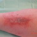
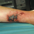
The examination of the wound allows identifying the origin of the sting: if there are several stings by several thorns, they are distributed at equal distance. Every sting presents a small wound with clear edges from 1 to 2 mm in length with an inflammatory area and a bluish halo (Fig. 40.3) and sometimes a small blood and fluid flow.
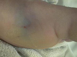
Fig. 40.3
Typical aspect of a sting with punctiforme lesion and bluish halo
Often the delay is more important and thus the inflammation has further extended with oedema and phlyctenas around the wound.
The association of a sudden, intense pain and these local aspects allows evoking the diagnosis of sting by stonefish.
40.2.6 Severity of the Wound Depends on Several Factors
The size of the fish: the bigger the stonefish is, the more venom is injected and the more serious is the wound.
The wound: the number and the depth of the stings.
The victim: his (or her) age, weight and medical history.
The time period until medical care is given.
40.2.7 Medical Complications
Presence of a foreign body which will have to be removed: a piece of thorn that would have broken into the wound. Medical imaging might be necessary if there is a doubt, though this is rare for the Synanceia verrucosa thorns are very resistant.
Related posts:


Stay updated, free articles. Join our Telegram channel

Full access? Get Clinical Tree


