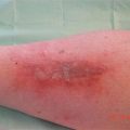
Fig. 43.1
Renecrosis after amputation on a compromised vascularisation. Result three month later after flammacerium
Indeed the guidelines will specify that before any debridement of the distal limb segments, particularly below the knee, the patient should be evaluated on the arterial side, and an ABPI should be realised combined to an arterial echo Doppler [24].
The guidelines also mention that the debridement is contraindicated in the presence of a dry gangrene or an ischaemic dry wound until a complete vascular evaluation is realised [21] and when waiting for a revascularisation procedure, the necrosis should be maintained as dry as possible in order to prevent infection.
43.4.2 Heel Pressure Ulcer
Following recommendations, arteriopathy prevention should include the detection of the early signs of vascular insufficiency in patients presenting risks of pressure ulcers aged less than 65 years and systematically over 65 years [25] (Fig 43.2).
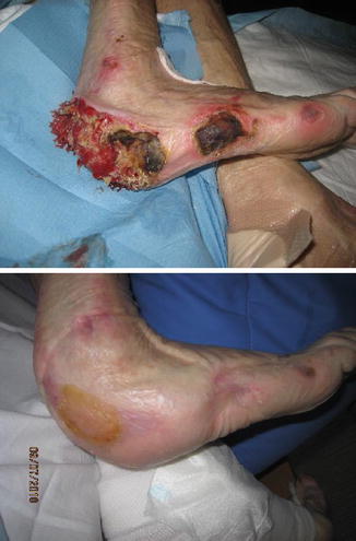
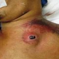
Fig. 43.2
Renecrosis of a pressure ulcer of the heel on a poorly nutrished arteriopathic patient. Results after 5 months of flammacerium
A cohort review driven by S.Meaume in 2007 established the links between arteriopathy and heel pressure ulcer [26]. Strategies for limiting the pressure over the heel are becoming insufficient to prevent PU due to the increase in rate of arteriopathy (most of the time unknown) in aged patients.
And reversely patients having heel PU may present associated risk factors like peripheral vascular disease leading to delayed wound healing [26, 27].
The vascular disease is a comorbidity associated with lower limb extremity PU [28], which should be recognised by analysis of the clinical evaluation, presence of pulses, time of capillary recolouration, oedema or mobility.
The realisation of an ABPI and its value >0.86 aim to eliminate an arterial disease in the PU pathogenesis [29].
Patients presenting an ABPI <0.8 should be considered as at risk of post heel PU debridement complication [30].
The procedure should be delayed if ABPI cannot be realised or if the values are low. A local strategy of stabilisation of the lesions should then be proposed before a procedure of revascularisation has been realised, if possible. In a recent article on NPUAP recommendations, caution would be to maintain as long as the possible a dry eschar over the wound (mummification) to prevent infection and odour [31].
43.4.3 The Diabetic Foot
Diabetic foot ulcers are most of the time contemporary with severe arteriopathy, out of the possibility of revascularisation, with a high risk of necrosis and severe infection, rendering this situation at the highest risk of non-traumatic amputation [32–34]. The stabilisation of necrosis is needed to save the limb (Fig 43.3).
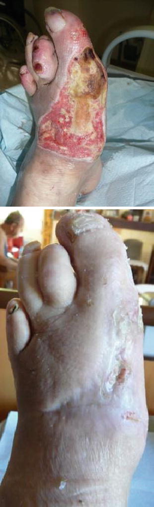
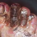
Fig. 43.3
Ischaemic diabetic foot ulcer
43.4.4 Cancer Wound
The debridement of a tumour wound is associated with a high risk of bleeding, pain and potentially a dissemination of cancerous cells. The wound bed is composed of necrotic and cancerous tissues due to the cancer, and it is impossible in most of the cases to remove all of them, healing being possible only if the carcinologic excision has been completed [35].
The necrosis is linked to the ischaemia of the tumour. It is so the local consequence and not the cause of a local disease (primitive cancer) or disseminated (skin metastasis) most of the time evolutive. The fact of leaving a wound covered with necrotic tissue is out of the normal recommended practice over a tumour wound, contrary to other types of chronic wounds. However the presence of prenecrotic tissue exposes the patient to infection, to the extension of the lesion and numerous inconveniences like heavy exudates, sévère odours linked to the présence of anaerobes [36] the management should then be aimed at complete healing but to clean the wound or to treat sequentially the symptoms like odours [37].
43.4.5 Osteoradionecrosis
One of the late complications of radiotherapy is skin radionecrosis. When standard care is not satisfactory (anti-inflammatory drugs, local care) [38] surgical excision and coverage surgical technique (skin grafting, flap) can be proposed. The complete excision is sometimes very difficult because of the anatomical structures involved and the poor quality of the covering skin. When the radionecrotic process involves bones in areas like the thorax or skull, it is sometimes impossible to extensively remove the bones and the underlying structures like the pleura or meningeal sheets. RMI or scanner is useful to check the bone involvement and the extent on the surrounding tissues. The decision to keep and stabilise the necrotic tissue using flmamacerium may be proposed when the skin is of poor quality.
43.4.6 Other Types of Wounds
Other situations may present contraindication to debridement like necrosis secondary to antimitotic toxic drugs (Avastin, corticosteroids) or other pathologies like sclerodermia where the pathology is linked to microvascular lesions. In these cases stopping the treatment or treating the cause may lead to wound healing. In some situations the treatment itself may lead to proper complications like the corticosteroid treatment (Figs. 43.4 and 43.5).
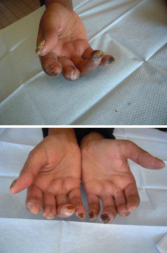
Fig. 43.4
Sclerodermia complications at the level of the distal end of the fingers: a perfect indication for flammacerium
Fig. 43.5
Long term successful management of a necrotic foot using flmamacerium (one year of treatment)
A waiting option should be opted aiming to gain time for better wound healing conditions, like the decrease of inflammation and pain which is always observed when using the Flammacerium cream.
43.5 Discussion and Perspectives
A retrospective study was realised and published in 2010 [39] to determine the efficacy of Flammacerium in stabilising the necrotic skin in chronic wounds. Ninety-nine patients were analysed, into which debridement was contraindicated (42 arterial leg ulcers, 30 PU, 5 cancer wounds, 10 trauma wounds and 12 others). This analysis showed a reduction in pain, exudates, odour and a positive effect on the quality of life, corresponding to an increase in patient comfort, social activity and psychological welfare. Moreover, when limiting the necrotic process, the use of the device extends the time of preparation of the surgical procedure.
The comfort offered by the Flammacerium, more than a protective crust, is due to the limitation of inflammation and pain, especially in arteriopathic patients, in osteoradionecrosis. The anticancer potential of the cerium compound still needs to be studied [4].
43.6 Conclusion
Flammacerium® has for a long time demonstrated its capacity in the prevention of severe infections in burns and its role in the inflammatory response. Its main advantages include its facility of application and use, allowing a mechanical barrier against infection. These properties may be extended to other pathologies like arteriopathies and all types of wounds where skin necrosis is present and debridement contraindicated.
Flammacerium has for a long period of time shown its efficacy in managing third degree burns. The main interest include an easy application, drastically abolishing pain at dressing change and pain induced by persistent inflammation. The obtention of a crust limit the possibility for germs to penetrate the wound, issuing to a better resistance to infection. The Flammacerium should certainly be proposed in other clinical indications, like chronic wounds when debridement is contraindicated. When a vascular impairment is in cause, the absence of elimination fold on the peripheral aspect of the wound covered with flammacerium is a benefit , limiting the surgical debridement, and allowing a revascularisation procedure in a non infected area. This product, even if debridement remains the first choice in most of the wounds, flammacerium may find a specific place in severely devascularised wounds.
Related posts:


Stay updated, free articles. Join our Telegram channel

Full access? Get Clinical Tree


