Chapter 37 Skin signs of gastrointestinal disease
It is logical that many diseases of the skin also involve the oral and anal mucosa because, embryologically, the foregut (forming the oral epithelium) and the hindgut (creating the anal mucosa) share a common ectodermal component in the first few weeks of fetal development. Therefore, the skin becomes a “mirror” of underlying pathology, both obvious and occult, in the gastrointestinal (GI) system.
Fitzpatrick JE: Cutaneous manifestations of gastrointestinal disease. In McNally PR, editor: Gastroenterology secrets, ed 3, Philadelphia, 2006, Elsevier Mosby, pp 559–566.
Kliegman R, Nelson WE: Nelson textbook of pediatrics. ed 18. Philadelphia, 2007, Saunders Elsevier, p 3147.
Do not forget about exogenous plant pigments, metabolic diseases, and trematode ingestion when examining a patient who appears jaundiced. The differential diagnosis of jaundice includes carotenemia (excessive ingestion of carotenoids), lycopenia (via tomato juice), Clonorchis sinensis (travel to Asia) or Fasciola hepatica (ingesting watercress) infection, and the sallow skin of myxedema.
Jaundice | Purpura |
Pigment changes | Loss of body hair |
Spider angioma | Gynecomastia |
Palmar erythema | Peripheral edema |
Dilated abdominal wall veins | Non-palpable gallbladder |
Hepatobiliary diseases are associated with alterations of the vasculature, including spider angiomas, palmar erythema, and cutaneous varices. Spider angiomas are classically associated with chronic liver disease, yet may also be seen in pregnancy, oral contraceptive use, and in normal persons, especially children. The vascular spider consists of a coiled central arteriole with smaller vessels radiating outward like the legs of a spider. In chronic liver disease, they are numerous and are found on the face, neck, upper chest, hands, and forearms. “Liver palms” refers to the mottled erythema and increased warmth of the palms (and sometimes the soles of the feet) in chronic liver disease. Palmar erythema also may be seen in pregnancy, lupus erythematous, pulmonary disease, and hyperthyroidism.
Portal venous hypertension due to chronic liver disease leads to the development of collateral circulation, with esophageal varices as an example. In the skin, this is observed as dilation of the abdominal wall veins (Fig. 37-1). Caput medusa refers to the dilated periumbilical veins and has been known for centuries as a marker of advanced liver disease. In men with chronic liver disease, induction of a “hyperestrogen state” (due to a decreased efficacy of estrogen breakdown in the liver) leads to gynecomastia, testicular atrophy; loss of axillary, truncal, and pubic hair; and a female pattern of pubic hair. Purpura, ecchymoses, and gingival bleeding reflect impaired hepatic production of various clotting factors, especially the vitamin K–dependent factors. Peripheral edema and ascites indicate hypoalbuminemia and/or portal venous hypertension.

Figure 37-1. Profile view showing dilation of the umbilical vein in the epigastrium. Enlarged abdominal wall veins reflect portal venous hypertension. The patient also had esophageal varices.
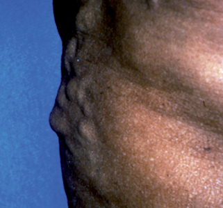
Table 37-1. Conditions Associated with GI Bleeding and Skin Lesions
INFLAMMATORY CONDITIONS | VASCULAR MALFORMATIONS AND TUMORS |
---|---|
Ulcerative colitisCrohn’s diseaseHenoch-Schönlein purpuraPolyarteritis nodosa | Hereditary hemorrhagic telangiectasia (Osler-Weber-Rendu)Kaposi’s sarcomaBlue rubber bleb nevus syndrome |
HEREDITARY POLYPOSIS SYNDROMES | MISCELLANEOUS |
Gardner’s syndromePeutz-Jeghers syndromeMultiple hamartoma syndrome (Cowden’s syndrome) | Ehlers-Danlos syndromePseudoxanthoma elastica |

Figure 37-2. Pyoderma gangrenosum. Large necrotic undermined ulcer of the lower leg with exposed tendon in a patient with ulcerative colitis.
(Courtesy of the Fitzsimons Army Medical Center teaching files.)
The exact cause of PG is unknown, but immune complex–mediated neutrophilic vascular reactions in the skin have been postulated. After the diagnosis of PG is made, the next step should be to look for an underlying cause. Important conditions to search for include chronic infectious hepatitis, inflammatory bowel disease (ulcerative colitis or Crohn’s disease), rheumatoid arthritis, lupus erythematosus, HIV infection, and leukemia. About 2% of patients with ulcerative colitis have PG, and the course of both illnesses maybe parallel. Some patients may have PG for several years before developing inflammatory bowel disease. In general, firstline treatment is corticosteroids and cyclosporine. Infliximab (TNF-α antibody) is the treatment of choice for PG with underlying inflammatory bowel disease or rheumatoid arthritis.
Brooklyn T, Dunnill G, Probert C: Diagnosis and treatment of pyoderma gangrenosum, BMJ 333(7560):181–184, 2006.

Stay updated, free articles. Join our Telegram channel

Full access? Get Clinical Tree

