MoorLDI flux image (1) predicts the dark and light blue areas as HP > 21. As shown in the clinical photographs, below, some healing occurred by post-burn day 20; these areas correspond to the areas of high flux on the moorLDI image (1). A biopsy was taken from the area indicated by moorLDI as HP > 21, prior to surgery, on post-burn day 20 (clinical photograph 7). The biopsy showed a third-degree burn wound.
Parts healed as predicted by HP type and the biopsy result was consistent with HP > 21 criteria.
Clinical Photographs
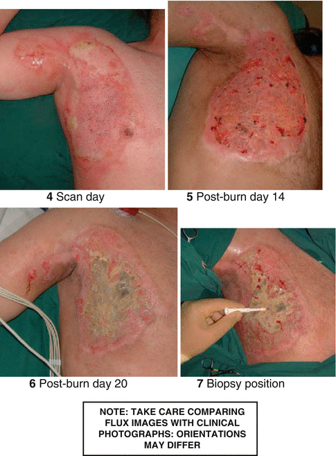
7.
Duplex scan
Duplex ultrasound is a special ultrasound technique that can assess how fast blood is flowing through a blood vessel. The test combines traditional ultrasound with Doppler ultrasound. Regular ultrasound uses sound waves that bounce off different structures of the body to create pictures. Doppler ultrasound records sound waves reflecting off moving objects, such as blood, to measure their speed and other aspects of how they flow. With addition of colour, the velocity can be measured. The accuracy and usefulness of Duplex ultrasound scanning in wound healing have been described in recent literature [5, 6].
8.
Thermography, that is, an imaging modality based on sensing heat emitted by a body, has the potential to image insults to skin including necrosis. It has been used to detect damage due to burns though its availability and acceptance have possibly been limited by the expense of the equipment and the instructor required to conduct the scans.
9.


CT angiogram/MR angiogram
Related posts:


Stay updated, free articles. Join our Telegram channel
Full access? Get Clinical Tree
