Skin changes in internal conditions
Skin signs are seen with many internal disorders and are not uncommonly their presenting feature. The astute dermatologist can recognize undiagnosed systemic disease.
Skin signs of endocrine and metabolic disease
Almost all endocrine diseases (and several metabolic defects) have cutaneous signs that depend on the over- or underproduction of a hormone or metabolite (Table 1).
Table 1 Skin signs of endocrine and metabolic disorders
| Disorders | Skin signs |
|---|---|
| Diabetes mellitus | Necrobiosis lipoidica, granuloma annulare, xanthomas, Candida albicans infection, ‘dermopathy’, neuropathic ulcers |
| Thyrotoxicosis | Pink soft skin, hyperhidrosis, alopecia, pigmentation, vitiligo, onycholysis, clubbing, pretibial myxoedema, palmar erythema |
| Myxoedema | Alopecia (including eyebrows), coarse hair, dry puffy yellowish skin (e.g. hands, face), asteatotic eczema, xanthomas |
| Addison’s disease | Pigmentation (p. 75), vitiligo, loss of axillary and pubic hair |
| Cushing’s disease | Pigmentation, hirsutism, striae, acne, obesity, buffalo ‘hump’ |
| Acromegaly | Thickened moist greasy skin, pigmentation, skin tags |
| Phenylketonuria | Fair hair and skin, atopic eczema (p. 36), photosensitivity |
| Hyperlipidaemia | Xanthomas (tuberous, tendinous, eruptive, plane), xanthelasma |
| Cutaneous porphyrias | Photosensitivity, blistering, skin fragility, atrophic scarring, thickening of skin, hypertrichosis, pigmentation (p. 46) |
Diabetes mellitus
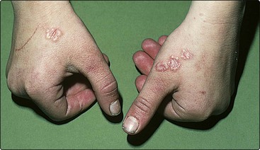
Candida albicans or bacterial infection is more common with untreated or poorly controlled diabetes. The neuropathy or arteriopathy of diabetes may result in ulcers on the feet (p. 73), and an associated secondary hyperlipidaemia can produce eruptive xanthomas (see Fig. 4). Diabetic dermopathy describes depressed pigmented scars on the shins, associated with diabetic microangiopathy. Necrobiosis lipoidica (Fig. 1), characterized by shiny atrophic yellowish–red plaques on the shins, was associated with diabetes in 65% of cases in one series, although others find a much lower figure. It affects less than 1% of all diabetics. Histologically, degenerate dermal collagen is seen with epithelioid cells and giant cells. The condition is chronic and may ulcerate. It is unresponsive to treatment. In contrast, granuloma annulare – recognized as palpable annular lesions on the hands, feet or face (Fig. 2) – is only rarely associated with diabetes and usually fades in 2 years. It must be differentiated from tinea corporis.
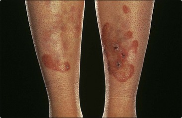
Fig. 1 Necrobiosis lipoidica.
Yellowish–red atrophic areas are seen on the shins of a diabetic patient.
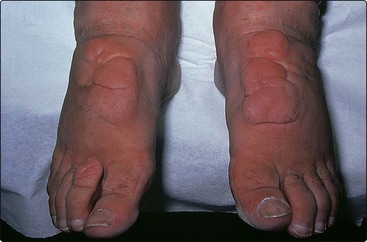
Thyroid disease
Both over- and underproduction of thyroxine result in skin and hair changes (see Table 1). Pretibial myxoedema (Fig. 3), seen in 1–10% of patients with hyperthyroidism, presents on the shins as raised erythematous plaques due to the deposition of mucin in the dermis. Topical steroids may be of benefit.